May 31, 2021
https://doi.org/10.23999/j.dtomp.2021.5.1
J Diagn Treat Oral Maxillofac Pathol 2021;57–66.
Under a Creative Commons license
HOW TO CITE THIS ARTICLE
Nosyr OV, Fesenko II, Khrulenko SI. Mandibular fractures: pre-operative panoramic radiography and duplication sign patterns. J Diagn Treat Oral Maxillofac Pathol 2021;5(5):57–66.
INSTITUTIONAL REPOSITORY
https://ir.kmu.edu.ua/handle/123456789/643
SUMMARY
The purpose of this essay is to present the multiple patterns of the duplication sign at the mandibular fracture line/gap visualized at the panoramic radiography as two-line fracture gap or pseudocomminuted fracture. We retrospectively reviewed the orthopantomography of patients with mandible fractures and presented nine patients with 12 duplication signs (also known as lambda course fracture line). On panoramic radiographs the fracture line/gap with duplication sign is visualized as two-line cortical bone discontinuity with or without dislocation due to the fact that the fracture gap runs asymmetrically on the external and internal cortical plates of the jaw. Knowledge of duplication sign patterns, artifacts is also crucial for the precise diagnosis and choice of correct management strategy.
INTRODUCTION
Panoramic radiography (PR) is a 2-dimensional zonography of all teeth, maxilla, mandible, and neighboring anatomical structures (like maxillary sinuses, zygomatic bones, hyoid, styloid processes, etc.).1 The history of the orthopantomography (ie, PR) begun at 1922 when the narrow-beam principle for jaw scanning was described by Zulauf in United States.2,3 Paatero (professor of clinical dental science) and Nieminen (engineer), both from Finland, were those whose close collaboration and multiple PR-related invented devices given the radiological and manufacturing world possibility to develop the modern orthopantomography. The term originally introduced by Paatero which means “orthoradial panoramic tomography”.3
During the last decades the PR equipment evolved from the conventional to digital systems which multiple advantages, like significantly lower radiation dose, quick usage of the images via different devices, etc., cannot be ignored not only by dentists of all specialties but also by oral and maxillofacial surgeons.4,5 Being financially affordable for the patients in Ukraine, PR starts to be a first line diagnostic tool in cases of traumatic injuries of the jaws.6,7 For example, in Kyiv, Ukrainian capital, the price for the digital PR varies from 190.74 UAH (ie, $6.82 USD) (in communal hospitals) to 280 UAH (ie, $10.02 USD) (in private diagnostic centers). Very often the doctors prefer PR over the X-rays in two different projections (en face [ie, posteroanterior] and lateral radiographic views).
Interpretation of the PR images in some cases in the patients with jaw fractures can be an uneasy task8 for the less experienced practitioners (interns, residents) due to such radiological signs like shadows’ superposition, duplication, presence of PR artifacts,9 and in case of the absence of radiologist`s conclusion.
This essay is the first English-language presentation of a multiple orthopantomography patterns of the duplication sign at the mandibular fracture lines/gaps.
MATERIALS AND METHODS
Panoramic images presented in this article were obtained at panoramic x-ray unit (Planmeca ProMax® 2D S3, Planmeca, Helsinki, Finland), Kyiv Regional Clinical Hospital by an experienced x-ray technician (S.I.K.; his experience – 25 years). This equipment was developed precisely for the maxillofacial imaging. The digital processing of radiographs was carried out using the Romexis Viewer software. The PR images were retrospectively analyzed. X-ray sign of shadows` superposition10 (ie, bone fragments overlapping [double radiopacity]11) and comminuted fractures were differentiated from duplication sign.
Nine patients of the age varied from 23 to 45 years old with duplication sign at mandibular fractures on PR images were analyzed (Figs 1–9). In seven of nine fracture cases (ie, in 77.77 percent) the bilateral mandibular fracture was established. In three of nine patients (ie, in 33.33 percent) the bilateral mandibular fractures with duplication sign at each fracture site were noted. So, totally 12 patterns of duplication sign were investigated.
DUPLICATION SIGN
Duplication sign is a radiographical sign which creates a false impression of the presence of a comminuted mandible fracture due to the fact that the fracture gap runs asymmetrically on the external and internal cortical plates of the jaw.10
Nardi et al name duplication sign as a lambda course of the fracture line.1 Lambda (a Greek letter) has an uppercase (Λ) and lowercase (λ) variants what is completely consistent with the various X-ray patterns of fractures. Predominantly, it can be noted an inverted lambda letter fracture pattern. Describing duplication sign by Greek letters, it`s also possible to apply an omicron (Greek letter) fracture pattern (its uppercase variant [Ο]). It can be visualized on the panoramic radiographs as a description variant of duplication sign. And in the article of Nardi et al, the symphysis fracture with lowercase lambda course description is presented.1
However, in our opinion, it is more appropriate to use a unified name duplication sign rather than application of different letters.
Based on our nine PRs with 12 duplication sign cases, we can summarize the following:
- The duplication sign can be visualized on the PR at any fracture`s localization (ramus, angle, body, and symphysis).
- In case of multiple mandible fractures, the bilateral duplication sign (ie, duplication sign at each fracture site) can also be noted.
- Duplication sign can be noted in both types of fracture – with and without fragments displacement.

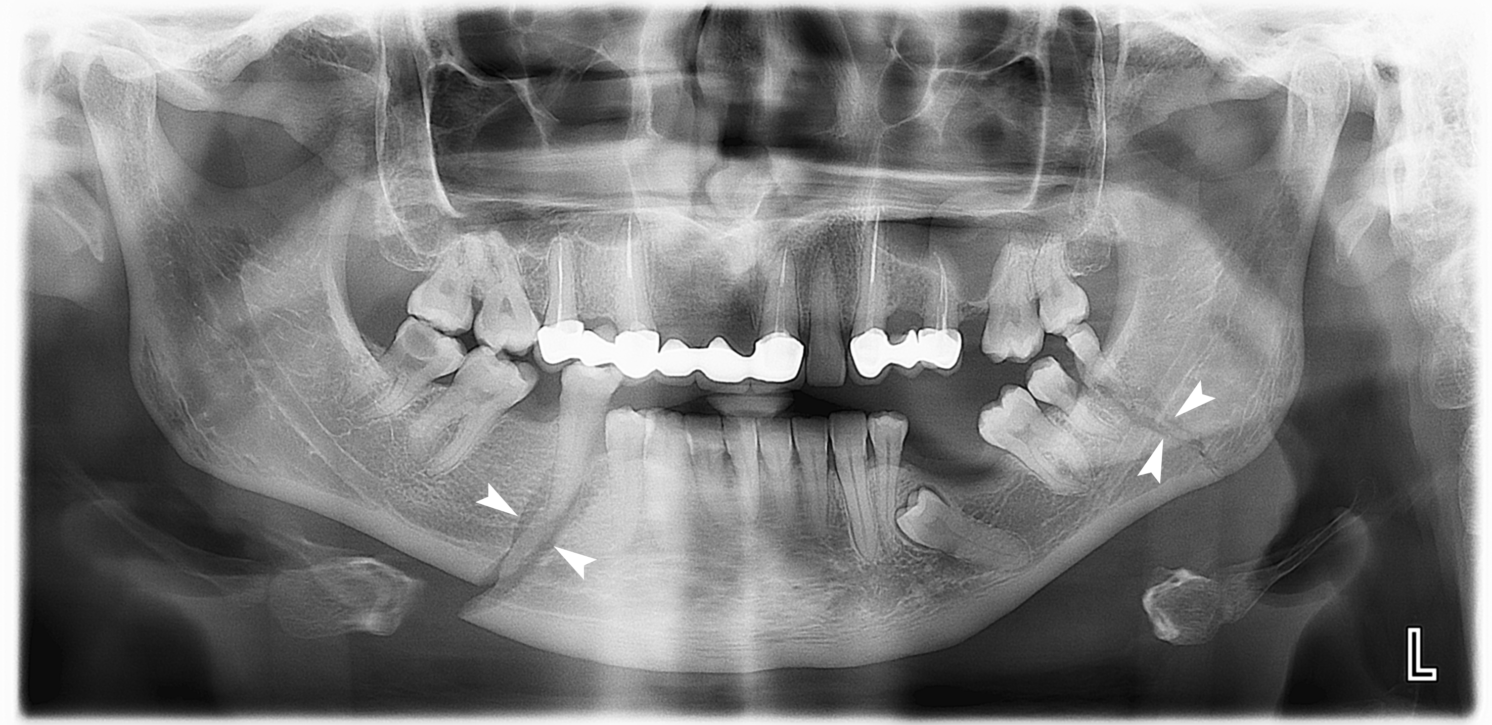
FIGURE 1. Case 1: A 34-year-old patient with bilateral mandibular fracture with duplication sign (arrowheads) at each fracture site.
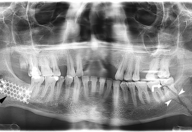
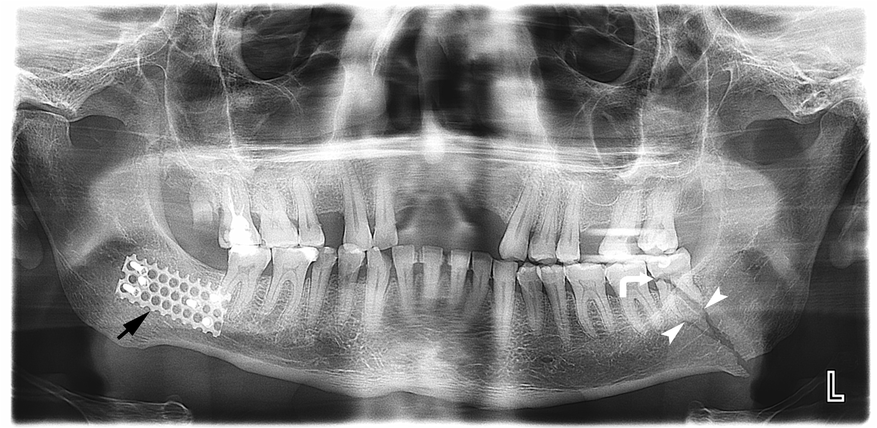
FIGURE 2. Case 2. A 39-year-old patient with mandibular fracture at the left angle with duplication sign (arrowheads) at the fracture gap. Curved arrow indicated on the fractured lower left third molar.
Osteosynthesis plate (arrow) in the area of consolidated fracture visualized at the right hemimandible.
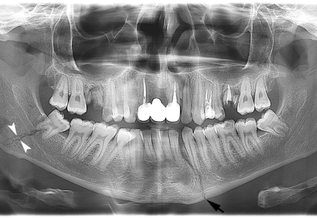
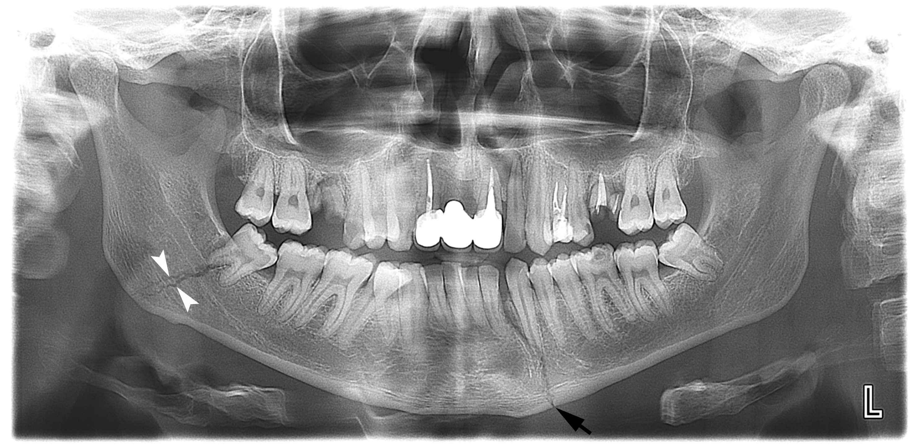
FIGURE 3. Case 3: A 23-year-old patient with bilateral mandibular fracture – at the right angle area with duplication sign (arrowheads) and one-line fracture (arrow) between tooth 3.2 and 3.3.
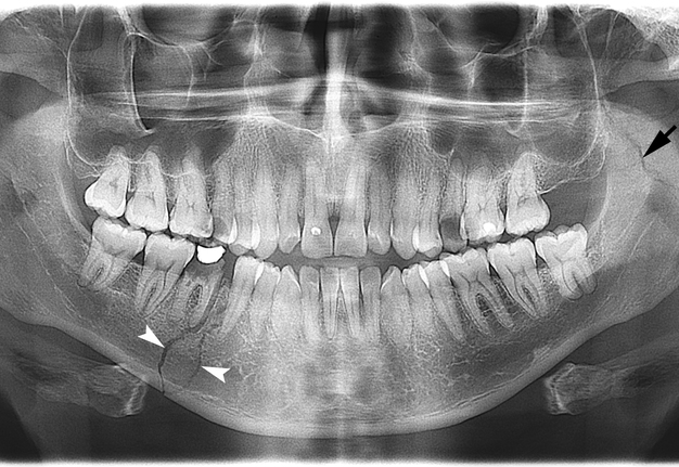
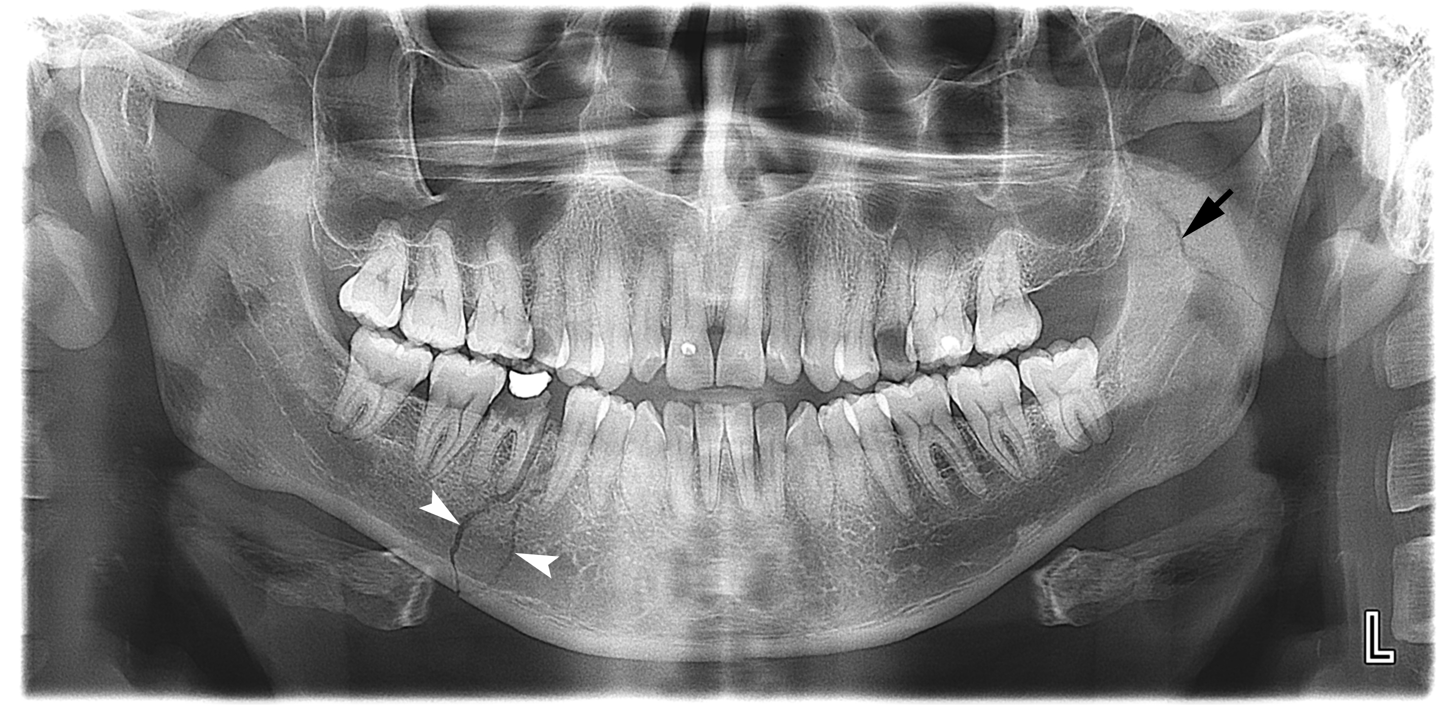
FIGURE 4. Case 4: A 33-year-old patient with bilateral mandibular fracture – at the right body area with duplication sign (arrowheads) and one-line fracture (arrow) of the left ramus.
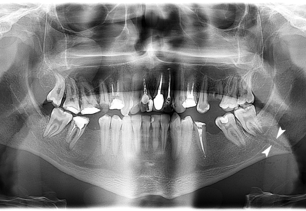
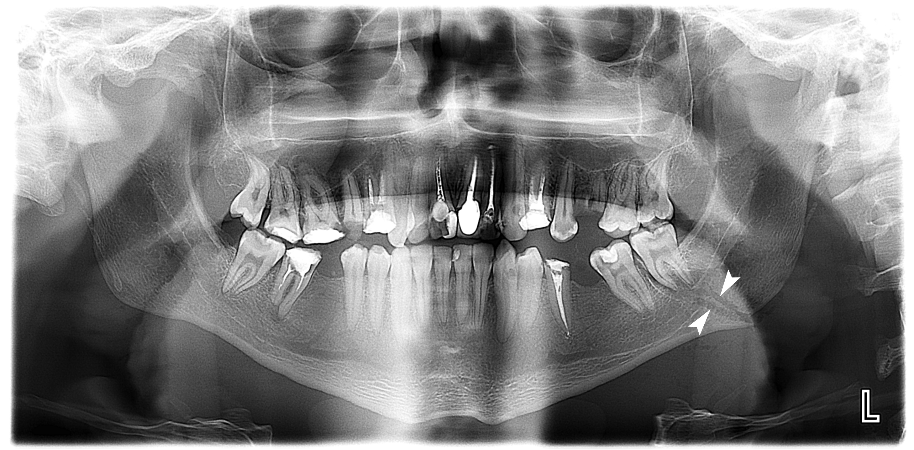
FIGURE 5. Case 5: A 38-year-old patient with unilateral mandibular fracture – at the left angle area with duplication sign (arrowheads).
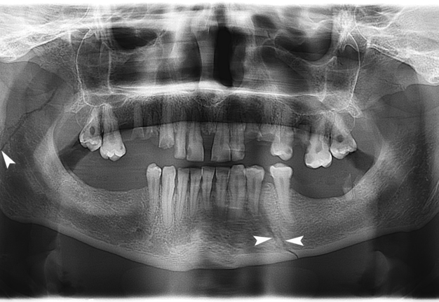
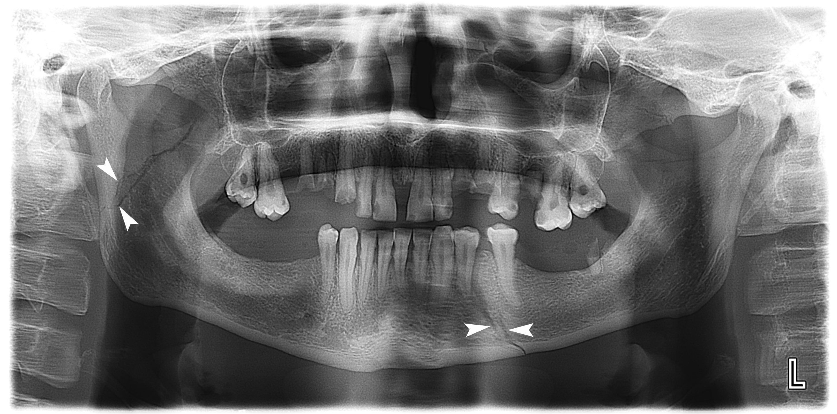
FIGURE 6. Case 6: A 25-year-old patient with bilateral mandibular fracture with duplication sign (arrowheads) at the right subcondylar fracture and at the
left body area.
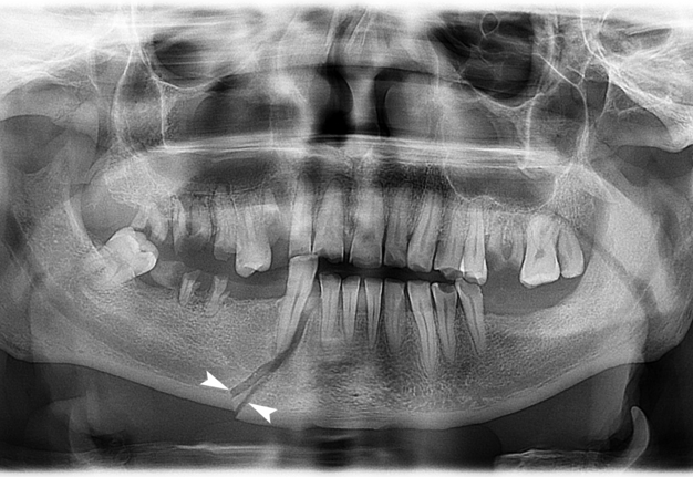
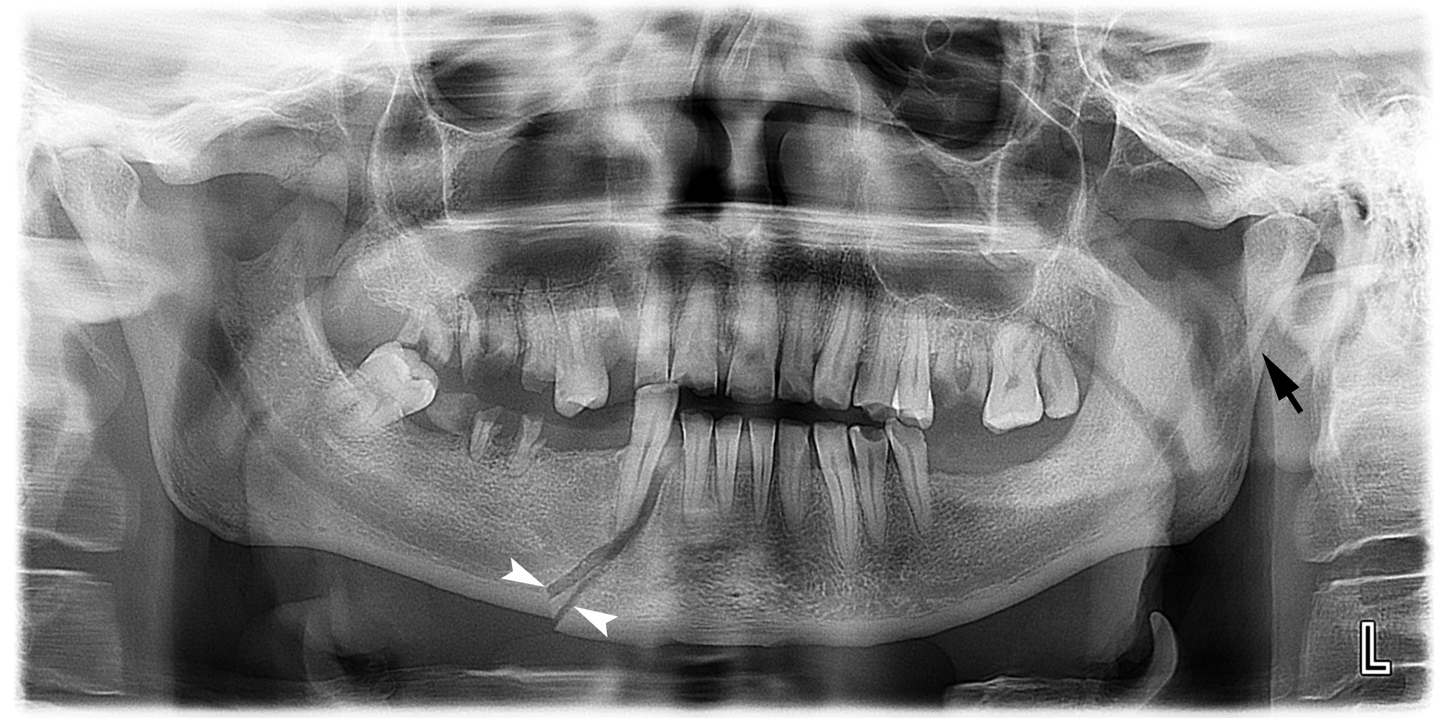
FIGURE 7. Case 7: A 43-year-old patient with bilateral mandibular fracture – at the right body area with duplication sign (arrowheads) and subcondylar
fracture (arrow) with shadows’ superposition sign.
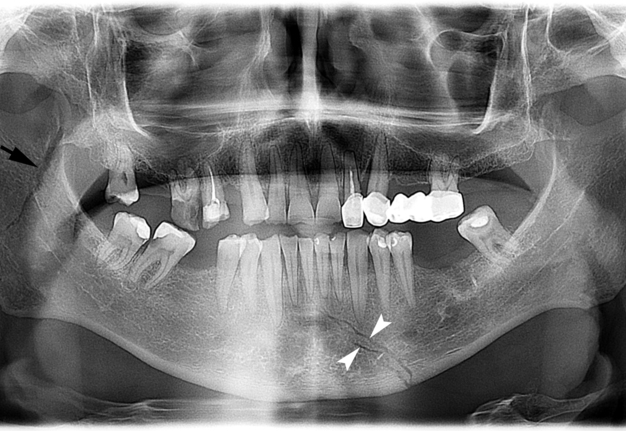
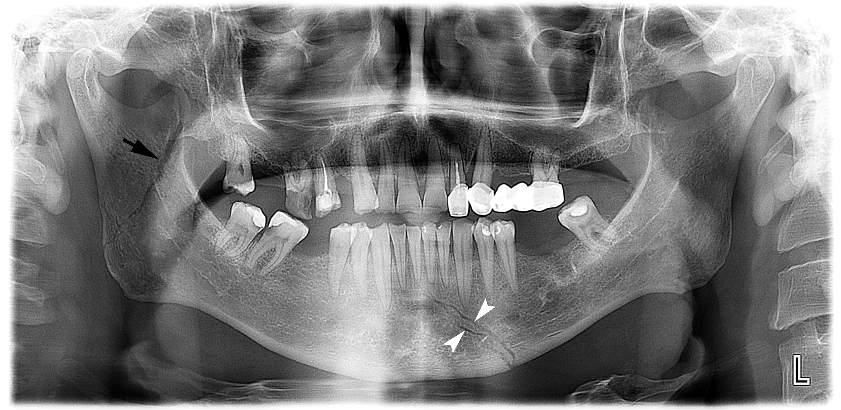
FIGURE 8. Case 8: A 45-year-old patient with bilateral mandibular fracture. The fracture area with duplication sign is indicated by arrowheads and one-line
fracture – by arrow.
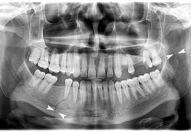
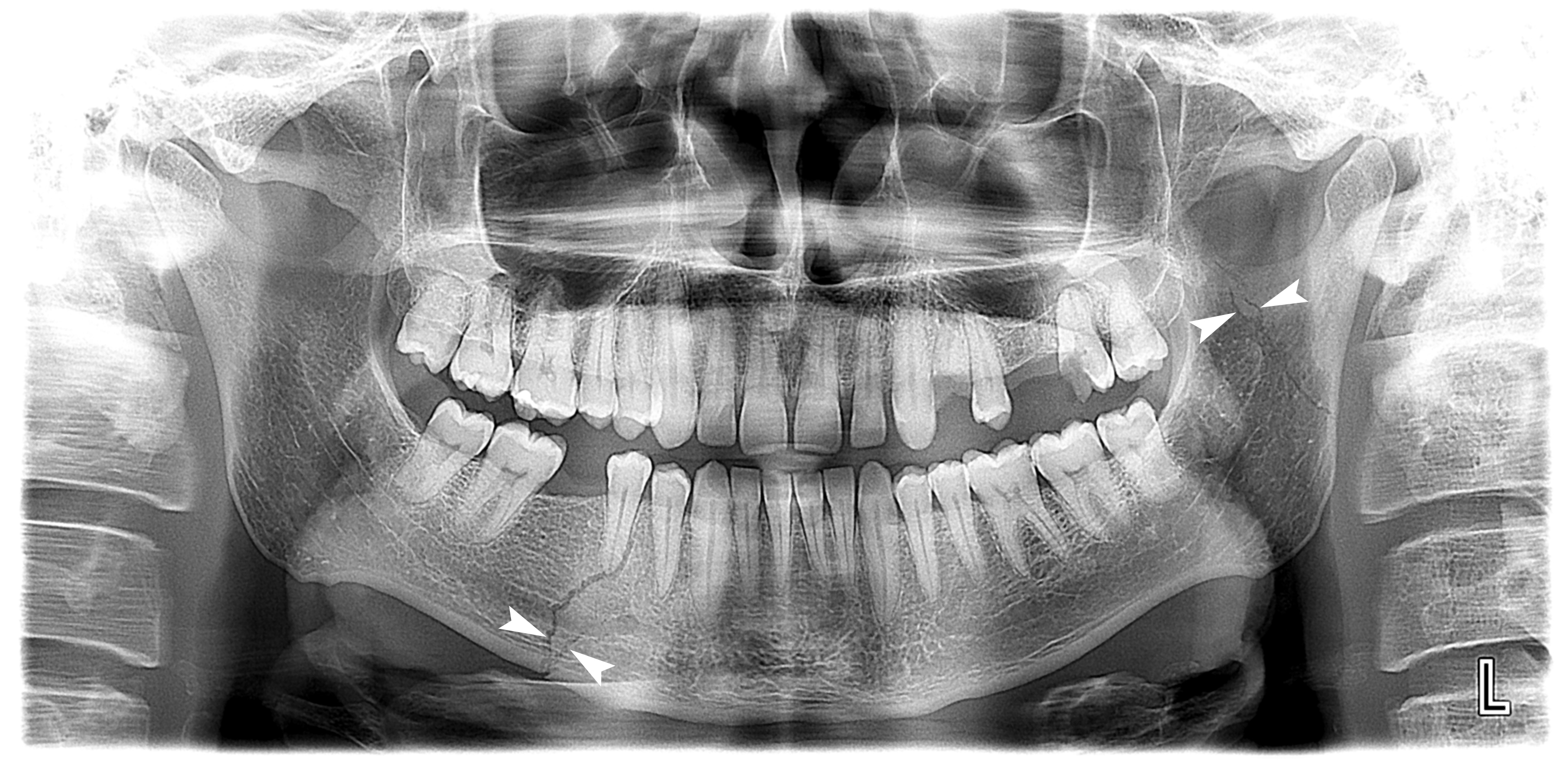
FIGURE 9. Case 9: A 36-year-old patient with bilateral mandibular fracture with duplication signs (arrowheads).
Of course, analysis and understanding of possible PR artifacts can also facilitate the establishment of the correct diagnosis.
Anyway, Albassal et al are right when emphasized on a need to perform a thorough clinical examination even when PR was done with a purpose not to miss a fracture site.8 Moreover, such clinical symptoms like indirect load (symptom of reflected pain), spatula symptom or a pressure on both mandibular angles can be helpful.10
Based on literature used in this article, five most common terms are widely used to describe panoramic radiography imaging and can be applied equally:
- Panoramic radiography.3,5,12–14
- Panoramic X-ray.8
- Panoramic dental radiography.4
- Orthopantomography (OPT15 or OPG16).
- Panoramic view.11
Also, only two terms were noted for the description of the same two-line fracture gap sign: duplication sign and lambda course.
CONCLUSIONS
Panoramic radiography is staying very affordable and helpful as an initial diagnostic tool in patient with mandibular and perimandibular tissues trauma. Knowledge of duplication sign patterns, artifacts is also crucial for the precise diagnosis and choice of correct management strategy.
AUTHOR CONTRIBUTIONS
Conceptualization: Nosyr OV. Data acquisition: Khrulenko SI. Data analysis, interpretation, and drafting of the manuscript: Nosyr OV. Approval of the final version of the manuscript: all authors.
REFERENCES
-
Nardi C, Vignoli C, Pietragalla M, Tonelli P, Calistri L, Franchi L, Preda L, Colagrande S. Imaging of mandibular fractures: a pictorial review. Insights Imaging 2020;11(1):30. Crossref | Medline | Google Scholar
-
Zulauf F. Panoramic XZ-ray apparatus. U.S. Patent No 1408559; 1922.
-
Hallikainen D. History of panoramic radiography. Acta Radiologica 1996;37(3):441–5. Crossref | Medline | Google Scholar
-
Sabarudin A, Tiau YJ. Image quality assessment in panoramic dental radiography: a comparative study between conventional and digital systems. Quant Imaging Med Surg 2013;3(1):43–8. Crossref | Medline | Google Scholar
-
Angelopoulos C, Bedard A, Katz JO, Karamanis S, Parissis N. Digital panoramic radiography: an overview. Semin Orthod 2004;10(3):194-203. Crossref | Google Scholar
-
Tymofieiev OO, Fesenko II, Tymofieiev OO. Condition of the teeth in fracture gap of the mandible. J Diagn Treat Oral Maxillofac Pathol 2017;1(1):41−53. Crossref | Google Scholar
-
Woldenberg Y, Gatot I, Bodner L. Iatrogenic mandibular fracture asso¬ciated with third molar removal. Can it be prevented? Med Oral Patol Oral Cir Bucal 2007;12(1):E70-2. Crossref | Medline | Google Scholar
-
Albassal A, Al-Khanati NM, Harfouch M. Could a digital panoramic X-ray not detect a displaced fracture of the mandible? Quant Imaging Med Surg 2021;11(8):3890–2. Crossref | Medline | Google Scholar
-
Izzetti R, Nisi M, Aringhieri G, Crocetti L, Graziani,F, Nardi C. Basic knowledge and new advances in panoramic radiography imaging techniques: a narrative review on what dentists and radiologists should know. Appl Sci 2021;11:7858. Crossref | Google Scholar
-
Tymofieiev OO. Manual of maxillofacial and oral surgery [Russian]. 5th ed. Kyiv, Ukraine: Chervona Ruta-Turs; 2012. Google Scholar
-
Larheim TA, Westesson P-L. Maxillofacial imaging. 1st ed. Berlin, Heidelberg, Germany: Springer-Verlag; 2006. Google Scholar
-
Sidorenko de Oliveira Capote T, de Almeida Gonçalves M, Gonçalves A, Gonçalves M. Panoramic radiography — diagnosis of relevant structures that might compromise oral and general health of the patient, emerging trends in oral health sciences and dentistry. In: Virdi M, editor. Emerging trends in oral health sciences and dentistry. IntechOpen; 2015. Crossref | Google Scholar
-
Rondon RH, Pereira YC, do Nascimento GC. Common positioning errors in panoramic radiography: a review. Imaging Sci Dent 2014;44(1):1–6. Crossref | Medline | Google Scholar
-
Farman AG, Kushner GM. Panoramic radiology in maxillofacial trauma. In: Farman AG, editor. Panoramic radiology. 1st ed. Berlin, Heidelberg, Germany: Springer-Verlag; 2007:155–66. Crossref | Google Scholar
-
Pandolfo I, Mazziotti S. Orthopantomography. 1st ed. Milan, Italy: Springer-Verlag Italia; 2013. Crossref | Google Scholar
-
Dimitroulis G. Temporomandibular joint disorders. In: Perry M, Holmes S, editors. Atlas of operative maxillofacial trauma surgery. 1st ed. London, United Kingdom: Springer-Verlag; 2020:753–80. Crossref | Google Scholar