October 26, 2022
J Diagn Treat Oral Maxillofac Pathol 2022;6: 131–4.
Under a Creative Commons license
HOW TO CITE THIS ARTICLE
Fesenko II. Odontogenic cutaneous fistula and abscess of the superficial peri-zygomatic area. J Diagn Treat Oral Maxillofac Pathol 2022;6(10):131–4. https://doi.org/10.23999/j.dtomp.2022.10.2
NATIONAL REPOSITORY OF ACADEMIC TEXTS
https://nrat.ukrintei.ua/en/searchdoc/2022U000198/
INSTITUTIONAL REPOSITORY
https://ir.kmu.edu.ua/handle/123456789/568
SUMMARY
Purulent processes of the zygomatic and peri-zygomatic area are not common. Among etiologies are: otitis media, complication of zygomatic implantation, osteomyelitis, medication-related osteonecrosis of the jaw, drug-related osteonecrosis, and odontogenic infection. This article highlights the first literature reported case of clinical presentation of odontogenic abscess and cutaneous fistula of the superficial peri-zygomatic area from the upper third molar. The preoperative and follow-up photographs of a 76-year-old Caucasian male patient are demonstrated. The uniqueness of this case is that until now the upper third molars never been published as a source of abscesses of the peri-zygomatic area.
INTRODUCTION
Purulent processes of the zygomatic and peri-zygomatic area are not common. Among etiologies are: otitis media,1,2 complication of zygomatic implants placement,3,4 osteomyelitis,5 medication-related osteonecrosis of the jaw (MRONJ), “Krokodil” drug-related osteonecrosis,6 and odontogenic infection7,8. The last one in the peri-zygomatic area can manifest in a form of sinus tract7, abscess8, and even phlegmon8. To our literature search in English language there is no publication data which presents the clinical images of odontogenic cutaneous fistula and abscess of the peri-zygomatic area from upper third molar. That is why we are presenting the pre- and postoperative images of such rare case in a male patient.
CASE
A 76-year-old Caucasian male patient was referred to our hospital by doctor-stomatologist-surgeon from the stomatological polyclinic in Kyiv region. The patient appeared in the Center of Maxillofacial Surgery in 2016 at 10:18 p.m. with a significant swelling, erythema, and pyorrhea from cutaneous fistula in a right peri-zygomatic area (Figs 1A and 2A). According to patient complaints the pain appeared in the tooth 1.8 (upper right third molar) a week before, and then the swelling in a right peri-zygomatic area appeared and began to increase with gradual erythema of the skin above it. Tooth 1.8 was extracted by doctor in a Kyiv region on the day of the visit to the polyclinic (i.e., out-patient clinic). Mouth opening was free. Panoramic radiography (Planmeca ProMax® 2D S3, Planmeca, Helsinki, Finland) performed in Kyiv Regional Clinical Hospital showed the empty socket of the tooth 1.8 and no radiological evidence of tooth remains.
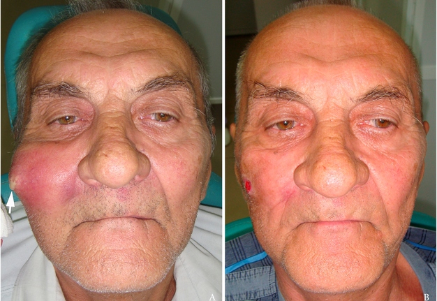
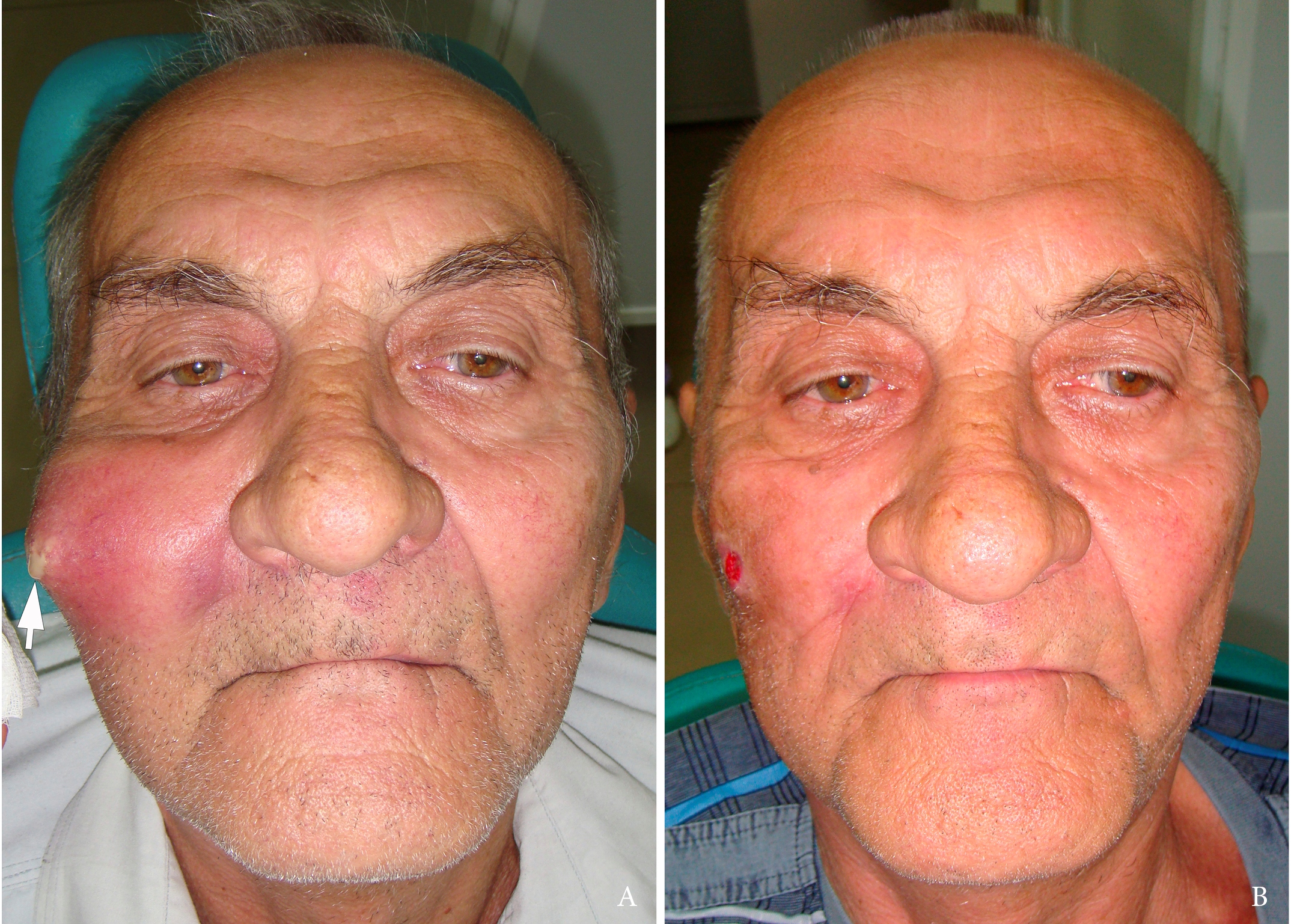
FIGURE 1. A, Preoperative view of a 76-year-old male patient with facial swelling in the right peri-zygomatic area. A notable purulent discharge from a skin fistula is labeled by arrow. B, Postoperative view on a day 4. The wound is healing by secondary intention.
Under the intramuscular injection of Dexalgini Inject 50 mg/2.0 ml (Laboratorios Menarini S.A., Barcelona, Spain; A. Menarini Manufacturing Logistics and Services Srl, Prato, Italy), first, applying the halsted-mosquito atraumatic hemostat (curved) the evacuation of the purulent content was performed. Then, finger revision of the purulent wound by a little finger was done. Purulent content in amount of ~6.0 ml was obtained. Irrigation of the wound with antiseptic solution was done and a sterile gauze bandage with Oflocain-Darnitsa® (Pharmaceutical Firm Darnitsa, PRJSC, Kyiv, Ukraine) was applied (inside the wound). There was no need to drain a purulent wound using tubular perforated drainage due to the evacuation of purulent contents through an expanded fistula.
General state and condition of the local tissues improved day by day. On a day 4 of the postoperative period, the wound healed by secondary intention (Figs 1B and 2B) and its condition was acceptable. The patient was discharged that day from the hospital with postoperative recommendations―Solcoseryl® ointment (MEDA Pharmaceuticals Switzerland GmbH, Dübendorf, Switzerland) for several days and carrying out dental prosthetics.
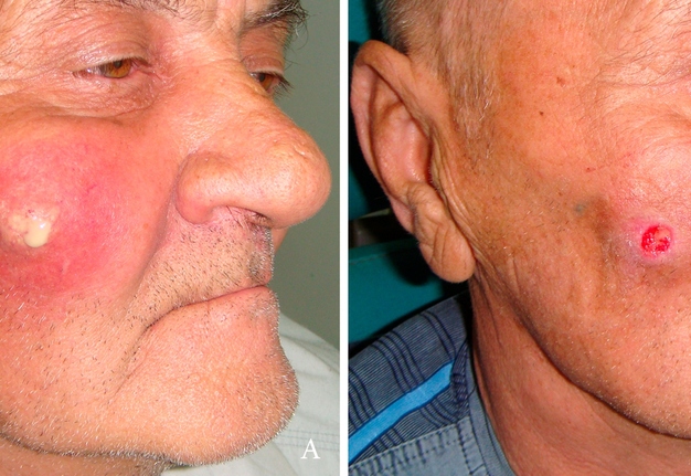
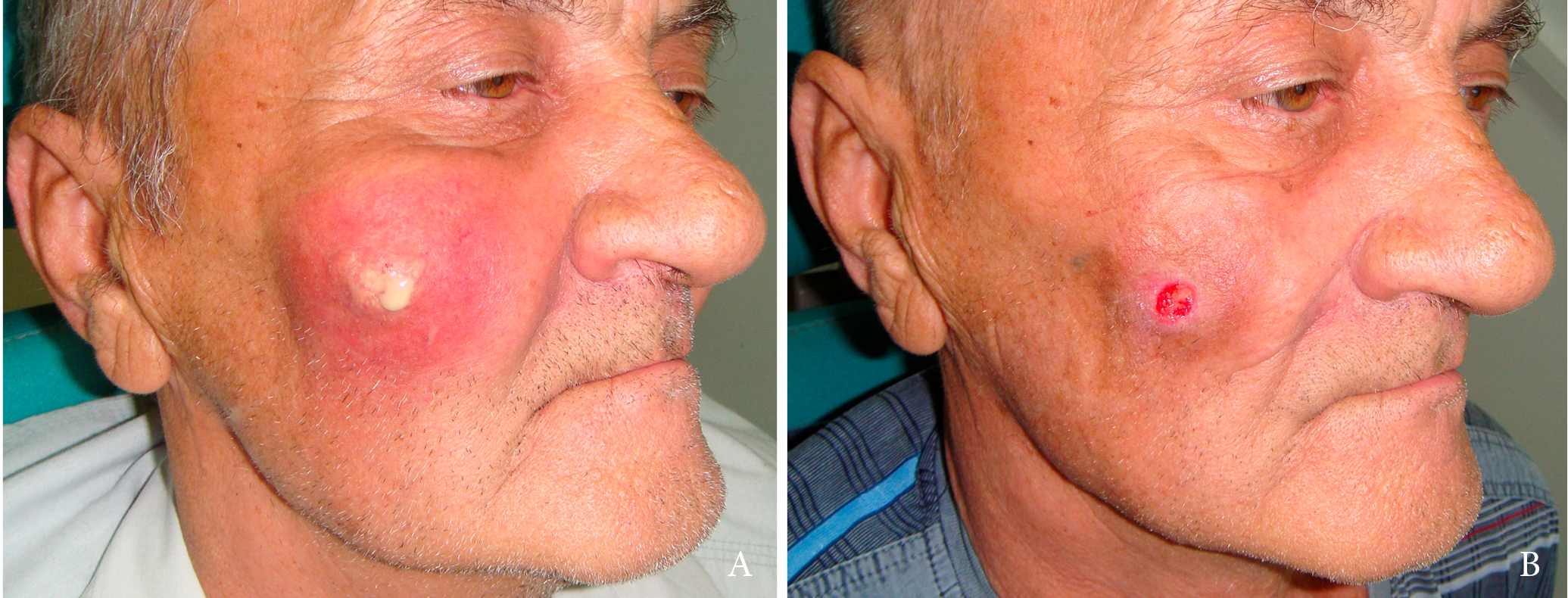
FIGURE 2. Preoperative (A) and postoperative view on a day 4 (B).
DISCUSSION
The term “zygomatic abscess” in English literature is commonly applied to the zygomatic root abscess―a rare extra cranial complication of acute otitis media.1,2 In the East European states (e.g., Ukraine), in oral and maxillofacial surgery publications, the term “zygomatic abscess/phlegmon” is applied to the purulent infection of superficial peri-zygomatic area.8 But only rare literature sources report odontogenic infection of such abscesses as the possible etiology.8 Reported odontogenic source is infection from the upper premolars or first molar.8
Peri-zygomatic (i.e., peri-malar) area includes the soft tissues which surround the zygomatic bone outside the orbit. To be clear, it’s reasonable to distinct superficial and deep peri-zygomatic area. Mendelson et al (2012) describe in the superficial peri-zygomatic region location of the prezygomatic cellular space.9
Odontogenic cutaneous sinus tract with skin fistula as one of its manifestations in the peri-zygomatic area have been reported, but the area was termed differently. Lee et al (2016) describe it as buccal cheek area.7 According to their data, in that area the odontogenic cutaneous sinus tract was manifested in 14.7 percent of odontogenic sinus tract cases (from upper first and second premolars [20 percent] and first and second molars [80 percent])7.
Suppurated congenital cysts of the zygomatic area are also reported and should be taken into account for the differential diagnosis in similar manifestations.8
Before this inflammatory process, the patient did not notice the presence of the skin fistula or swelling in this area. This fact and the absence of firm intraoral submucosal strand upon palpation allowed us to rule out the diagnosis of the odontogenic cutaneous sinus tract manifested with cutaneous fistula (Baba et al, 2017)10 or abscess. But this purulent process could be considered as one of the stages of sinus tract formation if the tooth was preserved without endodontic treatment.
Such purulent condition in that area should be managed as soon as possible to avoid zygomatic bone osteomyelitis, further spread of the inflammatory process, and long-term impairment of social life due to cosmetic problems.
CONCLUSIONS
This paper highlights the first literature reported case of clinical presentation of odontogenic abscess and cutaneous fistula of the superficial peri-zygomatic area from the upper third molar. The preoperative and follow-up photographs are demonstrated. The uniqueness of this case is that until now the upper third molars never been published as a source of abscesses of the peri-zygomatic area.
FUNDINGS
No funding was received for this study.
PATIENT CONSENT
The patient provided written consent for the use of his image.
REFERENCES (10)
-
Sathe N. Zygomatic abscess as a complication of otitis media. Natl J Maxillofac Surg 2011;2(2):181–3. Crossref
-
Kuczkowski J, Narozny W, Stankiewicz C, Mikaszewski B, Izycka-Swieszewska E. Zygomatic abscess with temporal myositis - a rare extracranial complication of acute otitis media. Int J Pediatr Otorhinolaryngol 2005;69(4):555–9. Crossref
-
Tzerbos F, Bountaniotis F, Theologie-Lygidakis N, Fakitsas D, Fakitsas I. Complications of zygomatic implants: our clinical experience with 4 cases. Acta Stomatol Croat 2016;50(3):251–7. Crossref
-
Vrielinck L, Moreno-Rabie C, Schepers S, Van Eyken P, Coucke W, Politis C. Peri-zygomatic infection associated with zygomatic implants: a retrospective longitudinal cohort study. Clin Oral Implants Res 2022;33(4):405–12. Crossref
-
Kang HJ, Lee JH, Kim YD, Byun JH, Shin SH, Kim UK, Jung IK. Osteomyelitis occuring in the zygoma caused by odontogenic maxillary sinusitis - case report [in Korean]. J Korean Assoc Oral Maxillofac Surg 2004;30(3):251–4.
-
Hakobyan K, Poghosyan Y. "Krokodil" drug-related osteonecrosis of midface: a case series. J Craniomaxillofac Surg 2019;47(12):1918–21. Crossref
-
Lee EY, Kang JY, Kim KW, Choi KH, Yoon TY, Lee JY. Clinical characteristics of odontogenic cutaneous fistulas. Ann Dermatol 2016;28(4):417–21.
-
Tymofieiev OO. Manual of maxillofacial and dental surgery [in Russian]. 5th ed. Kyiv: Chervona Ruta-Turs; 2012:276–318.
-
Mendelson BC, Muzaffar AR, Adams WP Jr. Surgical anatomy of the midcheek and malar mounds. Plast Reconstr Surg 2002;110(3):885–96; discussion 897–911. Crossref
-
Baba A, Okuyama Y, Shibui T, Ojiri H. Odontogenic cutaneous fistula mimicking malignancy. Clin Case Rep 2017;5(5):723–4. Crossref