

Virtual Surgical Planning for Osteosynthesis in Pediatric Craniofacial Surgery: The New State of Art
Available online 20 July 2026
J Diagn Treat Oral Maxillofac Pathol 2026;7: 100320
DOI: https://doi.org/10.23999/j.dtomp.2026.7.100320
Under a Creative Commons license
HOW TO CITE THIS ARTICLE
Kilipiris EG, Alvarez EM, Horn F. Virtual surgical planning for osteosynthesis in pediatric craniofacial surgery: The new state of art. J Diagn Treat Oral Maxillofac Pathol. 2026;10(7):100320.

ABSTRACT
Over the last decade, rapid advances in three-dimensional digital technologies have revolutionized the daily practice of pediatric craniofacial surgery, in general, and craniosynostosis surgery, more specifically. As with any new technological tool, they have been welcomed by pediatric craniofacial teams, who have found several useful applications in open craniosynostosis surgery, such as modeling craniofacial osteotomies. In this review, we analyzed the implementation of computer-assisted virtual planning in open cranial vault reconstruction for the surgical management of children with craniosynostosis, sometimes with the use of intraoperative navigation. Additionally, we documented the most common applications of virtual surgical planning technology in open craniosynostosis surgery, including modeling craniofacial osteotomies and manufacturing patient-specific biodegradable implants. We described the specific surgical workflow based on the virtual planning process. Finally, we presented some advantages and some limitations of these technological tools.
KEY WORDS
Craniosynostosis, virtual surgical planning, pediatric craniofacial surgery, craniofacial osteotomies, patient-specific biodegradable implants, resorbable osteosynthesis
INTRODUCTION
Pediatric craniomaxillofacial surgery involves comprehensive restitution of form, function, and aesthetics while treating dysmorphologies and defects. The anatomy is complex, and the limited blood reserve requires efficient, precise interventions. The topographic relations and compartments among anatomic structures are critically important for bony reconstruction. Planning such intricate surgery preoperatively is paramount, ensuring that geometric and quantitative factors, particularly those related to the craniofacial skeleton, are evaluated and appropriately accounted before the operation [1].
In recent years, advances in three-dimensional virtual simulation, digital imaging technology, and software have opened the door to a range of surgical planning techniques. Digital medical imaging technologies provide a basis for personalized surgery by applying surgical procedures to the individual patient before surgery. The earliest uses of 3D technology involved 3D-printed anatomical models of bone for hands-on visualization, diagnosis, and treatment planning. In recent years, technology has continued to evolve in driving personalized surgery. This is done using virtual tools translated for the pediatric operating room via model outputs, guides, templates, and 3D-printed implants. Now, customized implants can be made using 3D-printed models and templates, even tailored to a specific patient and surgical plan. Custom surgical guides and templates are 3D-printed from virtual surgical planning prior to surgery.
DISCUSSION
Digital Planning in Pediatric Craniomaxillofacial Surgery
Three-dimensional printing of implantable osteosynthesis plates is becoming increasingly common, with applications in several specialties. These osteosynthesis implants meet the same standards as their traditionally manufactured counterparts. This new area is known as virtual surgical planning. Virtual surgical planning is quickly becoming the standard of care in craniomaxillofacial surgery, especially in applications involving osteotomies and osteosynthesis. This also includes pediatric craniofacial reconstruction (Fig 1).
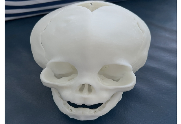
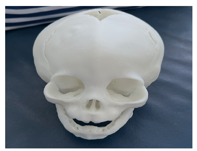
FIGURE 1. Stereolithographic model of an infant with metopic craniosynostosis.
Virtual surgical planning technology can also be leveraged to produce patient-specific implants. For example, simulated preoperative anatomy can be used in Computer-aided design (CAD)/Computer-aided manufacturing (CAM) production of customized reconstruction plates, such as bioabsorbable osteosynthesis plates that match the virtual surgical plan. In this application, osteotomy guides can also incorporate predictive hole placement, corresponding to screw hole locations in the final plate design, with bone depth maps provided intraoperatively to aid in screw length selection. This method often requires software and engineering skills to manipulate data accurately and efficiently over time.
A clinical transfer from the digital to the physical world must occur, typically using 3D printing technologies to translate the virtual surgical planning to the operating room. Transfer to the operating room can take place by creating bone-borne templates to guide osteotomies, templates to guide the position of the bone, or an advanced technique of predictive hole placement to guide the positioning of osteosynthesis plates.
Digital osteotomies can be transferred to the patient practically by designing a bone-borne guide. A bone-borne osteotomy guide is specifically designed to fit in one and only one location on the patient's anatomy. This bone-borne portion allows the guide to fit into the anatomy like a lock-in-key, thus forcing the guide and subsequent osteotomy into the digitally planned position. When the guide is positioned on the patient's anatomy as designed, the surgeon can perform the osteotomy with confidence that it is in the same position as discussed during the virtual surgical planning meeting.
Computer-Assisted Virtual Planning in the Surgical Correction of Craniosynostosis
Cranial vault reconstruction aims to provide a permanent, stable, and structurally sound reconstruction that accurately reproduces the skull's three-dimensional shape and surface contour. However, when repairing the three-dimensional bony architecture of the pediatric craniofacial skeleton, the craniofacial team is visually limited by access and by the inability to achieve complete three-dimensional exposure of the desired surgical sites. The accurate restoration of normal skull shape by manual shaping is virtually impossible, particularly when defects are large or located in anatomical sites with complex surface topography [2]. Also, because of the nonlinear nature of the bone in the craniofacial skeleton, even a small degree of error can lead to poor outcomes. Using 3D imaging, craniosynostosis deformities can be analyzed, and appropriate reference planes for a planning procedure targeting facial-skeleton symmetry can be defined [3]. Osteotomies can be designed preoperatively, and bones can be virtually configured to achieve the desired shape and features (Fig 2). Autogenous reconstructions are preferred in the open surgical repair for craniosynostosis correction because further skeletal growth is anticipated. Autogenous craniofacial reconstructions may include osteotomy, in which a facial skeletal segment is mobilized, reshaped, translocated, and fixated in a new position.
Despite the potential benefits and reproducibility of surgical outcomes, the use of CAD/CAM for cranial reconstruction in children with craniosynostosis has been reported in small numbers. Computer-aided design was first used for craniosynostosis repair in 1996, consisting of a 3D interactive mathematical model of the patient's skull that enabled preoperative procedural planning. Computer-aided manufacturing was introduced in 2011, when a craniofacial team digitized 103 pediatric skulls from normocephalic children, averaged the surfaces within specific age groups (8-12 months), and manufactured customized stainless steel templates of the fronto-orbital region of the normative infant head. Then they used these templates to aid intraoperative fabrication of the supraorbital bandeau and the bandeau plates used in fronto-orbital remodeling to treat unicoronal, bicoronal, or metopic synostosis.
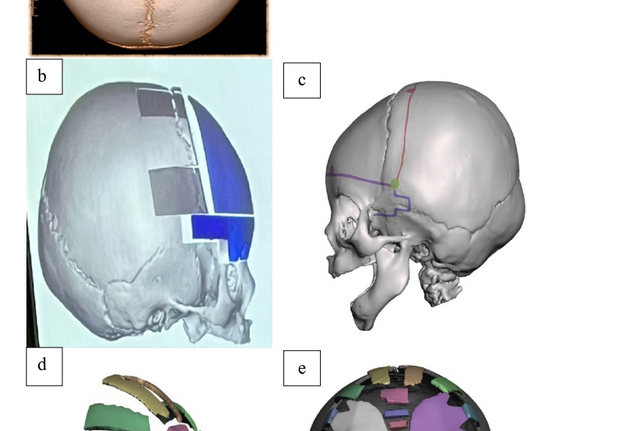
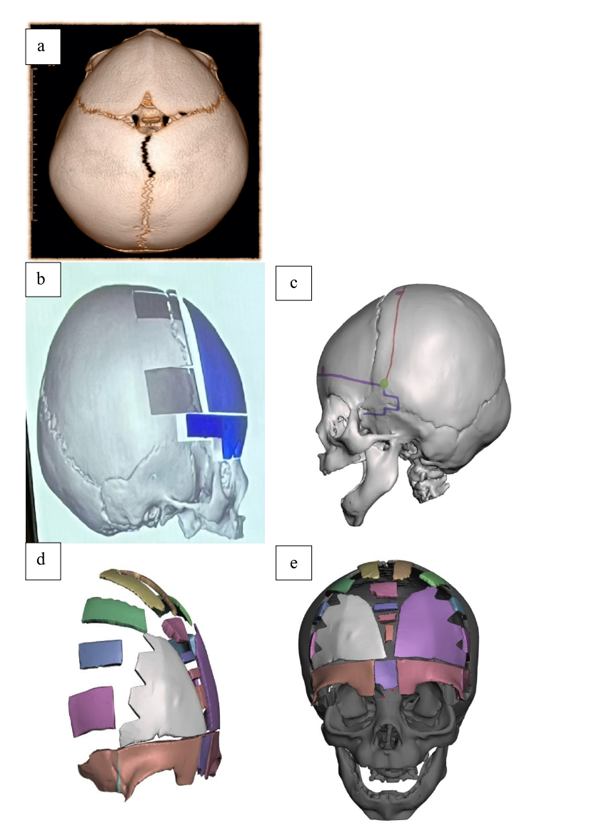
FIGURE 2. (a) A 3D CT reconstruction of a 9-month-old child with trigonocephaly as a result of metopic suture synostosis. (b-c). Different osteotomy techniques are designed preoperatively. (d-e) Projected postoperative state with a wider endocranial bifrontal angle and advancement of the fronto-orbital bandeau.
Applications in Open Cranial Vault Reconstruction
1. Modeling Craniofacial Osteotomies
The surgical correction of craniosynostosis involves cutting and reshaping the bones of the calvarium to achieve a more normal head shape, commonly occurring between 6 and 12 months of age. Upper facial osteotomies can be extremely complex. They rely not only on a transposition of a major anatomical segment but also on a reshaping or reconfiguration of the osteotomy segment itself. This generally necessitates multiple simultaneous osteotomies. Modeling these osteotomies is, therefore, a challenge, and reliably implementing them in the operating room is particularly difficult [4]. A simpler and more effective approach is to only model the final desired morphological result and then manufacture an intraoperative template of this ultimate contour. All the intervening steps (i.e., specific placement of osteotomies, the number of osteotomy segments, and the direction and degree of transposition of individual segments) are performed manually by the surgeon at the time of surgery. The surgeon's objective is to cut and mobilize osteotomy segments to fit the computer-generated template. This technique of prereconstructed templates for osteotomy and bone rearrangement has been effectively employed in the reconstruction of congenital deformities. It provides improved precision in osteotomy design, enabling better reconfiguration of the skull and facial bones. In addition, the osteotomies are performed with appropriate angles to allow optimal bone-bone contact and, therefore, optimal bone healing.
2. Manufacturing Patient-Specific Biodegradable Implants
Virtual surgical planning technology can also be leveraged to produce patient-specific implants [5]. Simulated preoperative anatomy can be used in the CAD/CAM production of various alloplastic implants, such as bioabsorbable osteosynthesis plates. For example, if fronto-orbital advancement is indicated, the degree of surgical advancement and contouring of the fronto-orbital complex is determined primarily by the surgeon's experience, as reliable data are currently unavailable. This is due to challenges to the three-dimensional measurement of the initial bony position and, consequently, the surgical outcome and the individual growth patterns of patients.
Although the transfer of digital planning using osteotomy guides and non-resorbable implants is no longer a technical challenge, the transfer into pediatric surgery has been limited to date to the use of osteotomy guides [6]. Customized reconstruction plates can be produced with CAD/CAM to match a virtual surgical plan. In this application, osteotomy guides can also incorporate predictive hole placement, corresponding to screw hole locations in the final plate design, with bone depth maps provided intraoperatively to aid in screw length selection.
The preoperative CT scan is processed to allow 3D osteotomies for a standard fronto-orbital advancement procedure. Drill holes are marked before the simulation. Two different types of drill holes are required: i) for fixation of the mobile segments and ii) for reliable 3D orientation of the resorbable patient-specific implant. Based on symmetry, the segments are moved and aligned with the ideal outer contour. Once the virtual planning is complete, intersections between adjacent segments are closed to allow for computer-assisted design and construction of the resorbable patient-specific implant. The previously marked drill holes can now be integrated into implant construction with a 1.6 mm diameter. This allows for the use of self-retaining screws. After that, the construction can be transferred to the plating system company, and the resorbable patient-specific implant can be built.
Intraoperatively, osteotomies are performed according to the cutting guide, and the bony segments are fixed with the bioresorbable patient-specific implant and resorbable screws. Internal fixation with resorbable plates and screws remains the gold standard, and individually shaped 3D resorbable implants are now commercially available. Test models show no significant discrepancy between computer-assisted construction and a resorbable patient-specific implant, with the drill holes, shape, and extent being congruent. The specific design of the resorbable implant, with two types of drill holes, allows predictable positioning. First, the bony segments are repositioned to the implant during fronto-orbital advancement. Second, the implant is fixed to the immovable temporal bone, which is especially important for the sagittal and vertical dimensions. The application of bioresorbable patient-specific implants to reassemble an osteosynthesis implant is time-saving [7].
Surgical Workflow Based on Virtual Surgical Planning
Using 3D planning software, a patient's skull can be compared to its age and size-matched counterpart. It generates templates for intraoperative calvarial osteotomy and remodeling, as well as positioning guides for accurate skull vault shaping [8]. For this reason, a recent craniomaxillofacial CT scan is required for virtual surgical planning in craniosynostosis correction. Due to the rapid growth during infancy, large changes in size and shape can occur in a relatively short time. This prohibits a considerable delay between scan acquisition and surgical correction using CT-based guides [9]. To avoid such morphological changes associated with growth and to provide suitable cutting guides during surgery, ideally, a high-resolution computed tomography scan of the patient's craniofacial skeleton with 3D reconstructions should be performed before surgery. Age-matched normative datasets developed for patients aged 8 to 12 months serve as a powerful reference tool during virtual surgical planning [10].
This 3D CT scan is used to plan the osteotomies and the placement of osteosynthesis plates with virtual surgical planning reconstruction. The images are sent to the biomedical engineering team in the Digital Imaging and Communications in Medicine (DICOM) format. The CT scan DICOM file is transferred to 3D cutting software, enabling segmentation of the cranio-orbital region and the creation of 3D planning models. Burr holes, osteotomy lines, and osteosynthesis plates are placed by the craniofacial surgeons on the virtual model, taking into account the patient's specific anatomy, avoiding critical anatomical structures such as the sagittal sinus and bone emissary veins, and selecting the ideal locations for osteosynthesis plates. Afterward, these planned cuts are virtually manipulated by superior and lateral expansion of bone segments flanking the center cut of a bone strip to match the dimensions of a normative cranium. Using this schematic, a model of the patient's skull is produced with a planned osteotomy overlay. Also, a stereolithographic model can be generated from the virtual surgical plan to allow surgeons to manually verify, in three dimensions, their simulation and the osteotomies [11]. Once the surgeons approve the stereolithographic models, the latter can be finalized. The 3D planning models are transferred to specialized software that generates files for printing the guides on a 3D printer. These are then used to render a sterilized cutting guide and a normative, age-specific child cranial template with outlines for the planned reconstruction [12]. The model, cutting guide, and template are created using 3D printing. The cutting and positioning guides created are sterilized for use in the operating room. Intraoperatively, the sterile manufactured cutting guide is used to mark the location of planned osteotomies on the skull. The burr holes are made with a round burr above the orbital bandeau and behind the coronal sutures, according to the cutting guide. Osteotomies are performed and positioned within the expanded outlines on the normative cranial template (Fig 3). The osteotomized bone segments of the skull are placed in the positioning guide. Thanks to this, the orbital bandeau is reshaped to achieve the correct angle, and the frontal bones are positioned and attached in the correct location, as established in the simulation, using resorbable plates and screws. The expanded top of the calvaria is placed and secured to the native bone.
Collaboration of the whole craniofacial surgical team is necessary for these complex reconstructions. All team specialists can attend the initial planning meeting to ensure the surgical plan is acceptable to all involved [13]. Planned osteotomy sites can be verified against planar CT slices to confirm safe distances from underlying intracranial anatomy, such as the sagittal and transverse dural venous sinuses [14].
For clinical transfer, a series of marking and positioning guides is used. After exposure of the calvarium, bone-borne marking guides are placed to mark all planned osteotomies and orientation characters. The craniofacial team will perform the osteotomies that define the overall craniotomy's outer borders, removing the affected calvarium as intact as possible. This premarked subtotal calvarium is then cut and reshaped on a back table, following the premarked cutting paths. Commonly, bone segments are moved, rotated, and reoriented in a complex way to achieve the final desired shape. To help facilitate these movements, the previously marked registration characters are used in conjunction with matching features in a set of positioning guides. These guides reinforce the appropriate positioning of these bony flaps prior to fixation. One common technique involves inserting all bony flaps into a bowl-shaped set of positioning guides before plating on the intracranial surface of the reshaped segments. The guides can then be removed, leaving a neocalvarium that can be replaced and fixated onto the skull base to cover the cranial defect. Fixation is then performed by using resorbable plating systems.
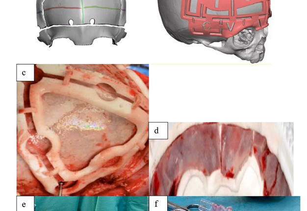
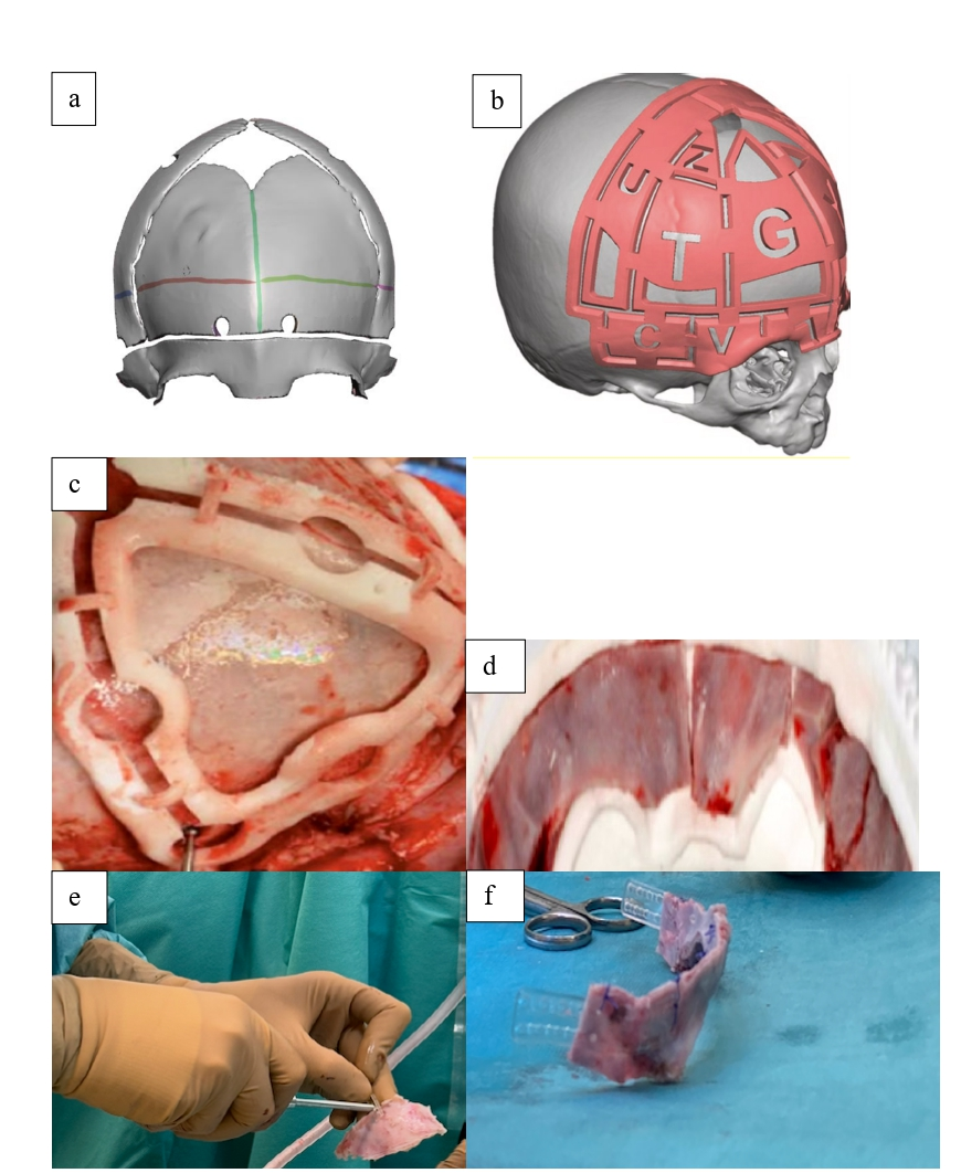
FIGURE 3. (a) Virtual bone configuration. (b) A cutting guide virtual simulation can accurately translate the surgical plan from the computer to the operating room for modeling craniofacial osteotomies through 3D-printed templates like this one for metopic synostosis repair. (c) Supero-lateral view of the trigonocepahlic skull showing the placement of the cutting guide on the frontal bone for making the burr holes and the osteotomies. (d-e) The cranial positioning guide receives the osteotomized and reconstructed fronto-orbital bandeau for osteosynthesis during the reconstructive phase of the fronto-orbital advancement. (f) The newly created banbeau is ready to be fixated in the accurate advanced position.
Intraoperative Navigation
Navigation has proven to be a valuable new tool for managing complex craniofacial cases, ensuring the correct repositioning of segments and immediate reconstruction with complex bone grafts and resorbable plates in 3D, in real time. With intraoperative navigation, advanced images can be obtained at the operative site. Intraoperative imaging increases the accuracy and safety of surgery by enabling real-time control of individual operative steps through accurate navigation [15]. A preoperatively acquired 3D reconstruction of CT images is constantly projected during the operation. A localizer tracks the patient's head position using fiducial markers placed on stable and reliable anatomical points. A surgical probe can then be used alone or mounted on the surgical instruments, which will be directly visualized in relation to the patient's anatomy (Fig 4). In specific cases, virtual planning can be combined with navigation guidance technology to achieve even greater accuracy.
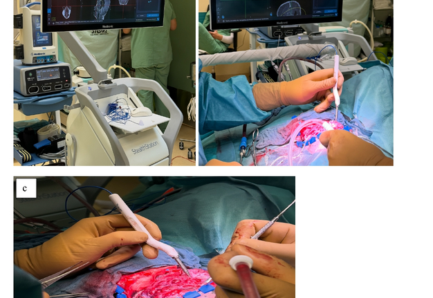
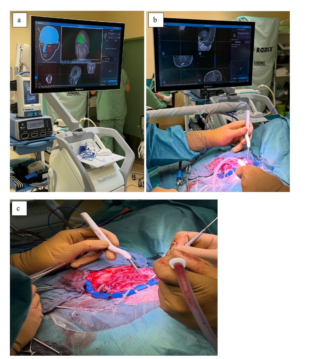
FIGURE 4. (a) Registration points are recorded using the tracked pointer tool on the skull of a 7-month old patient with Crouzon syndrome who underwent posterior cranial vault expansion. (b-c) A surgical probe is used to evaluate the optimal position before the accurate placement of the reconstructed bone segments and the resorbable osteosynthesis plates.
Virtual Surgical Planning Advantages in Craniosynostosis Surgery
3D computer-assisted surgical planning offers potential benefits by improving the accuracy and efficiency of the surgical procedure, leading to more reliable, predictable outcomes. Described benefits include enhanced parental understanding of surgical pathology and treatment plans, facilitated learning and training for surgical residents, with a significant decrease in the learning curve for performing complex operations, and streamlined requirements for surgical instruments [16]. In addition, it highlights the establishment of a safe and easy workflow. It provides a better understanding of the surgical procedure and surgical planning, leading to greater self-confidence.
These techniques are teachable, have a shallow learning curve, allow precise anatomic bony reconstruction, reduce operating time ranging from 45 to 90 minutes based on case complexity and the surgical team’s familiarity with the surgical guides. To quantify time savings in the operating room tracking incision-to-closure time and comparing it between Virtual Surgical Planning (VSP)-guided surgeries and standard, traditional, manual cranial vault cases is most commonly adopted. Furthermore, since VSP patient-specific surgical templates aim to minimize the time-consuming bone fragment remodeling and osteosynthesis, we can quantify the time savings using this specific timeline metric.
Furthermore, the recognized potential for efficient surgery, improved cosmetic outcomes, and decreased intraoperative stress with a displacement of time-consuming osteotomy and osteosynthesis planning to the preoperative setting, the development of practiced plans derived from multiple trial-and-error schematizations, and outcomes that precisely match preoperatively intended results, outweigh the limited downside of requiring a preoperative planning session.
VSP reduces surgical subjectivity and complications. The decrease in complications from craniosynostosis surgery can be quantified by measuring specific intraoperative, early postoperative, and late postoperative parameters. Intraoperative time reduction decreases the anesthetic exposure and blood loss. This would reduce the anesthetic risks exposure to the growing child and the costs associated with treatment [17]. Successful reduction of intraoperative blood loss can be easily quantified in milliliters and has been well documented in VSP patient groups compared with traditional groups.
A marked decrease in blood loss in VSP-guided craniosynostosis surgeries translates directly to reduced transfusion volumes. To quantify and accurately measure a decrease in blood transfusion requirements using VSP and 3D-printed surgical templates compared to traditional surgery, pediatric craniofacial units measure the calculated blood loss based on the patient’s pre- and post-operative hematocrit values and total blood volume, as the estimated blood loss is highly subjective. In addition, clinical studies can use transfusion volume and rate as metrics by comparing the absolute volume of packed red blood cells transfused during and after the surgical procedure.
When postoperative scans are used, volumetric and linear accuracy can be evaluated by measuring discrepancies between the planned 3D outcome and the postoperative scan results. Compared to conventional surgical and reconstructive methods, VSP has been shown to reduce linear discrepancies. Finally, based on revision rates, a reduction in complex secondary or late revision surgeries can indicate an improved surgical and reconstructive precision with the implementation of VSP.
It is also noted that parental expectations are better established through the provision of a tactile aid that models expected results, as well as improved safety profiles due to preoperatively navigated cuts. The surgery has already been performed once virtually, enabling multiple vantage points not present intraoperatively and early recognition of obstacles and interferences. In these conditions, 3D planning assists in the diagnosis, treatment simulation, surgical guide manufacture, and evaluation of the simulated surgical steps [18]. Because a digital surgical plan exists, there is, for the first time, an objective measure of surgical outcomes. This measure can not only help train the next generation but also establish a baseline for comparison among surgeons, centers, and geographic locations.
CAD/CAM Technology Limitations
The risks of surgical planning cannot be completely obviated. One common pitfall is that, in many instances, such a detailed presurgical plan does not allow for intraoperative decision-making when it references itself. It can also prohibit the surgeon from changing the surgical plan based on intraoperative findings. In addition, the use of CAD/CAM increases the overall time dedicated to the surgical procedure due to preoperative planning and validation of the virtual tools, and it also increases the overall procedure costs. CAD/CAM technologies involve investment in sophisticated software and hardware and, as a result, can be expensive to implement and use. Financial limitations are the most frequent barrier in institutions in resource-constrained areas. For this reason, the development of low-cost solutions, including the “in-house” production of surgical cutting guides using open-source software and “in-house” printers, and limited services from external companies, has gained traction and resulted in long-term cost savings.
Furthermore, using CAD/CAM technology necessarily requires modeling from a CT scan, with potential risks of radiation exposure. Moreover, it is essential to emphasize that although CAD/CAM virtual surgical planning is a precise and valuable tool, preoperative and intraoperative common sense should prevail. Virtual planning should be used as a guide and a supplement rather than as an absolute plan. Fine surgical adjustment and surgical judgment are still required.
CONCLUSION
This review showed that an optimized virtual surgical-guiding workflow enables greater surgical efficiency and individualized surgical precision in cranial vault reconstruction for craniosynostosis patients. However, these 3D printing-based techniques cannot replace a surgeon’s technical abilities and clinical judgment. Thus, future research must be directed towards rigorous assessment tools to determine whether the use of such technologies makes a significant difference, primarily in the patient’s outcome, in the craniofacial team’s work, and, more broadly, in the pediatric hospital's shared commitment, compared to conventional methods.
REFERENCES (18)
-
Bell RB. Computer planning and intraoperative navigation in cranio-maxillofacial surgery. Oral Maxillofac Surg Clin North Am. 2010;22(1):135-156. https://doi.org/10.1016/j.coms.2009.10.010
-
Tucker S, Cevidanes LHS, Styner M, et al. Comparison of actual surgical outcomes and 3-dimensional surgical simulations. J Oral Maxillofac Surg. 2010;68(10):2412-2421. https://doi.org/10.1016/j.joms.2009.09.058
-
LoPresti M, Daniels B, Buchanan EP, et al. Virtual surgical planning and 3D printing in repeat calvarial vault reconstruction for craniosynostosis: technical note. J Neurosurg Pediatr. 2017;19(4):490-494. https://doi.org/10.3171/2016.10.PEDS16301
-
Khechoyan DY, Saber NR, Burge J, et al. Surgical outcomes in craniosynostosis reconstruction: The use of prefabricated templates in cranial vault remodelling. J Plast Reconstr Aesthet Surg. 2014;67(1):9-16. https://doi.org/10.1016/j.bjps.2013.09.009
-
Essig H, Lindhorst D, Gander T, et al. Patient-specific biodegradable implant in pediatric craniofacial surgery. J Craniomaxillofac Surg. 2017;45(2):216-222. https://doi.org/10.1016/j.jcms.2016.11.015
-
Rana M, Chui CHK, Wagner M, et al. Increasing the accuracy of orbital reconstruction with selective laser-melted patient-specific implants combined with intraoperative navigation. J Oral Maxillofac Surg. 2015;73(6):1113-1118. https://doi.org/10.1016/j.joms.2015.02.014
-
Bell RB, Markiewicz MR. Computer-assisted planning, stereolithographic modeling, and intraoperative navigation for complex orbital reconstruction: a descriptive study in a preliminary cohort. J Oral Maxillofac Surg. 2009;67(12):2559-2570. https://doi.org/10.1016/j.joms.2009.07.098
-
Mardini S, Alsubaie S, Cayci C, et al. Three-dimensional preoperative virtual planning and template use for surgical correction of craniosynostosis. J Plast Reconstr Aesthet Surg. 2014;67(3):336-343. https://doi.org/10.1016/j.bjps.2013.11.004
-
Steinbacher DM. Three-dimensional analysis and surgical planning in craniomaxillofacial surgery. J Oral Maxillofac Surg. 2015;73(12 Suppl):S40-56. https://doi.org/10.1016/j.joms.2015.04.038
-
Saber NR, Phillips J, Looi T, et al. Generation of normative pediatric skull models for use in cranial vault remodeling procedures. Childs Nerv Syst. 2012 Mar;28(3):405-410. https://doi.org/10.1007/s00381-011-1630-7
-
Kobets AJ. Virtual modeling, stereolithography, and intraoperative CT guidance for the optimization of sagittal synostosis reconstruction: a technical note. Childs Nerv Syst. 2018 May;34(5):965-970. https://doi.org/10.1007/s00381-018-3746-5
-
Hassfeld S, Muhling J. Computer assisted oral and maxillofacial surgery-a review and an assessment of technology. Int J Oral Maxillofac Surg. 2001;30(1):2-13. https://doi.org/10.1054/ijom.2000.0024
-
Cevidanes L, Tucker S, Styner M, et al. 3D surgical simulation. Am J Orthod Dentofacial Orthop. 2010;138(3):361-371. https://doi.org/10.1016/j.ajodo.2009.08.026
-
Chauvel-Picard J, Beuriat PA, Daurade M, et al. Is computer-assisted design and manufacturing technology useful in the surgical management of trigonocephaly? J Craniomaxillofac Surg. 2021;49(11):993-999. https://doi.org/10.1016/j.jcms.2021.06.003
-
Luebbers H, Messmer P, Obwegeser JA, et al. Comparison of different registration methods for surgical navigation in cranio-maxillofacial surgery. J Craniomaxillofac Surg. 2008;36(2):109-116. https://doi.org/10.1016/j.jcms.2007.09.002
-
Seruya M, Borsuk DE, Khalifian S, et al. Computer-aided design and manufacturing in craniosynostosis surgery. J Craniofac Surg. 2013;24(4):1100-1105. https://doi.org/10.1097/scs.0b013e31828b7021
-
Xia JJ, Phillips CV, Gateno J, et al. Cost-effectiveness analysis for computer-aided surgical simulation in complex cranio-maxillofacial surgery. J Oral Maxillofac Surg. 2006;64(12):1780-1784. https://doi.org/10.1016/j.joms.2005.12.072
-
Xia JJ, Shevchenko L, Gateno J, et al. Outcome study of computer-aided surgical simulation in the treatment of patients with craniomaxillofacial deformities. J Oral Maxillofac Surg. 2011;69(7):2014-2024. https://doi.org/10.1016/j.joms.2011.02.018