

Management of Cephalohematoma: A Reference Center Experience and Description of a Refined Surgical Technique
Manuscript received 25 May 2026
Revised 27 July 2026
Accepted 28 July 2026
Available online 08 August 2026
J Diagn Treat Oral Maxillofac Pathol 2026;8: 100322
DOI: https://doi.org/10.23999/j.dtomp.2026.8.100322
Under a Creative Commons license
HOW TO CITE THIS ARTICLE
Vélez Restrepo D, Urrego González A, Salas Vargas JE. Management of cephalohematoma: A reference center experience and description of a refined surgical technique. J Diagn Treat Oral Maxillofac Pathol. 2026;10(8):100322.

ABSTRACT
Backround: Cephalohematoma is a common perinatal complication that typically resolves spontaneously. However, persistent cases may progress to calcification from the periphery toward the center of the hematoma after 4 weeks, resulting in cranial asymmetry and aesthetic deformity. There is currently no global consensus on the optimal timing and method of intervention. Methods: We analyzed the experience of a craniofacial reference center in Medellín, Colombia, in the management of both non-calcified and calcified cephalohematomas. Acute cases were initially observed. Persistent fluid collections were managed via percutaneous aspiration. Early “egg-shell” ossifications underwent minimally invasive removal, while mature calcified lesions with significant asymmetry were treated via craniotomy and tangential burring at approximately 12 months of age. Results: Twenty-two patients underwent percutaneous aspiration without complications or residual deformity. Seven patients with calcified cephalohematoma underwent surgical correction (mean age: 11 months). The mean operative time was 201 minutes, with a mean estimated blood loss (EBL) of 15 mL. No blood transfusions, infections, or reoperations were required. All patients achieved satisfactory aesthetic outcomes at 12-month follow-up. Conclusion: A staged management algorithm—ranging from aspiration to a specialized craniotomy and burring technique—provides safe, reproducible, and aesthetically satisfactory results for cephalohematoma at various stages of evolution.
KEY WORDS
Cephalohematoma, percutaneous drainage, calcification, craniectomy, cranial asymmetry
INTRODUCTION
Neonatal cephalohematoma is a subperiosteal hemorrhage confined by cranial sutures, with an estimated prevalence of 0.5% to 3% [1]. Risk factors such as vacuum extraction, occiput presentation, prematurity, fetal scalp electrode use, and gestational diabetes have been described.
While most cases resolve within the first weeks of life, complications such as infection, jaundice, anemia, and progressive calcification may occur [3]. Calcified cephalohematoma poses a significant challenge, as it leads to permanent cranial vault asymmetry and potential psychosocial impact [5].
Despite its prevalence, a standardized treatment algorithm remains elusive. Some authors suggest intervention for collections exceeding 50 mm after the first month of life to prevent ossification [4]. This study describes our institutional experience and a refined surgical technique for calcified lesions that prioritizes bone remodeling and minimizes the need for blood transfusions.
METHODS
Patient Selection and Evaluation
A retrospective review was conducted of patients referred to our craniofacial unit between September of 2022 to September of 2025. Parent informed consent was obtained before photographs were taken both pre and postoperative. This study was approved by the hospital ethics committee.
Neonates with large (diameter > 50 mm measured by computed tomography or ultrasound) [2] or bilateral cephalohematomas were monitored for 2-3 weeks. If the collection persisted or was associated with refractory hyperbilirubinemia, intervention was indicated. For calcified lesions, diagnosis was confirmed via low-dose cranial computed tomography (CT) with 3D reconstruction to plan the surgical approach.
Percutaneous Aspiration Technique
Aspiration was performed at the bedside under local anesthesia (topical EMLA [eutectic mixture of local anesthetics]). Under strict aseptic conditions, a 22-gauge IV catheter connected to a 20-mL syringe was used. Following drainage, a pressure dressing was applied for 24 hours (Fig 1).
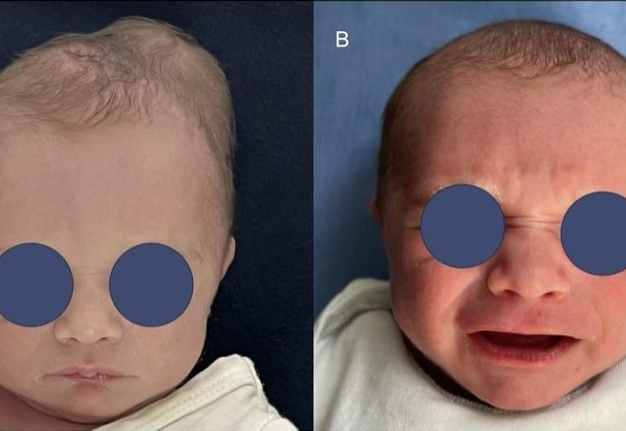
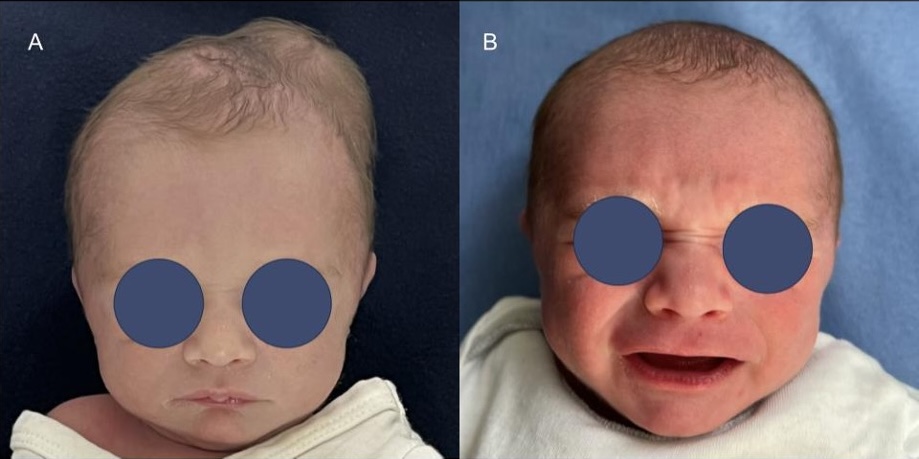
FIGURE 1. Figure demonstrates a preoperative (A) and postoperative (B) photo of a patient treated by percutaneous aspiration technique.
Minimally Invasive “Egg-Shell” Resection (Early Ossification Stage)
For patients in the early stage of ossification—characterized by a thin, “egg-shell” layer on CT—a minimally invasive approach is preferred. A 2-cm linear incision is made over the vertex (apex) of the cephalohematoma. Following subperiosteal elevation, the incipient ossified layer is fenestrated using a high-speed burr. The organized fibrous content and residual hematoma are then evacuated. The remaining calcified shell is meticulously resected using rongeurs. Finally, the bone margins are leveled with a bone rasp to ensure a smooth contour and a seamless transition with the surrounding calvarium. This technique minimizes surgical trauma and avoids the need for extensive craniotomies in early infancy. Figure 2 depicts an exemplary case in a 5 weeks old patient with bilateral early ossified cephalohematomas.
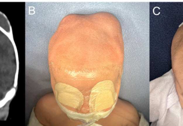
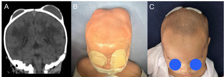
FIGURE 2. A five-week-old patient with bilateral early ossified cephalohematomas, treated with minimally invasive “egg-shell” resection. Computed tomography (CT) findings in coronal view (A). Pre-operative frontal view (B). First post-operative day frontal view (C).
Surgical Management of Calcified Lesions (Late Ossification Stage)
For mature calcified cephalohematomas, we employed a “Craniotomy and Translucency-Guided Burring” technique. Under general anesthesia, a wavy (stealth) incision was performed. The exostosis was demarcated using methylene blue. A craniectomy was performed using a 3-mm match-head burr and a pediatric craniotome. The key step involves tangential burring of the external cortex of the bone flap. The endpoint of burring was determined by bone translucency (transillumination) and manual palpation to ensure a uniform thickness comparable to the surrounding normal bone (Fig 3). The remodeled flap was then secured using a resorbable fixation system (Resorb X; Gebrüder Martin, KLS Martin Group), and standard closure with absorbable sutures was performed. Patients were admitted to the surgical ward and discharged home the following day.
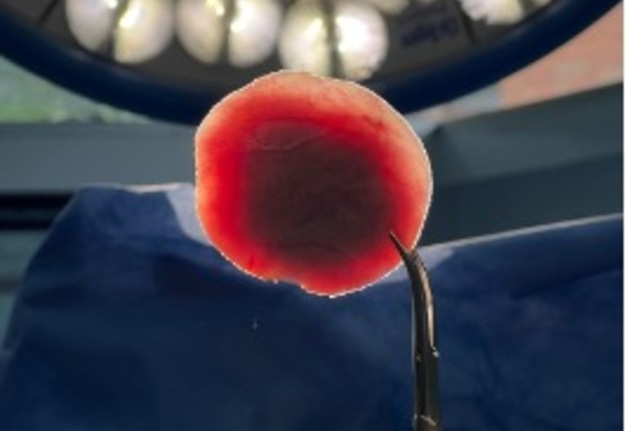
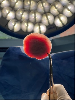
FIGURE 3. Typical translucency after craniectomy.
RESULTS
A total of 29 patients were treated. In the non-calcified cohort (n=22), percutaneous drainage successfully resolved all collections. In the surgical cohort (n=7), the mean age at surgery was 11 months (range 4–16 months) (Table 1). Two patients were operated using a minimally invasive approach (egg-shell resection) and 5 patients were operated using the “guided burring” technique. The mean operative time was 201 minutes, and estimated blood loss (EBL) was
remarkably low (mean: 15 mL), precluding the need for any blood transfusions. No postoperative complications, such as hematoma or surgical site infection (SSI), were observed. An exemplary case is depicted on Figure 4.
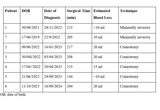
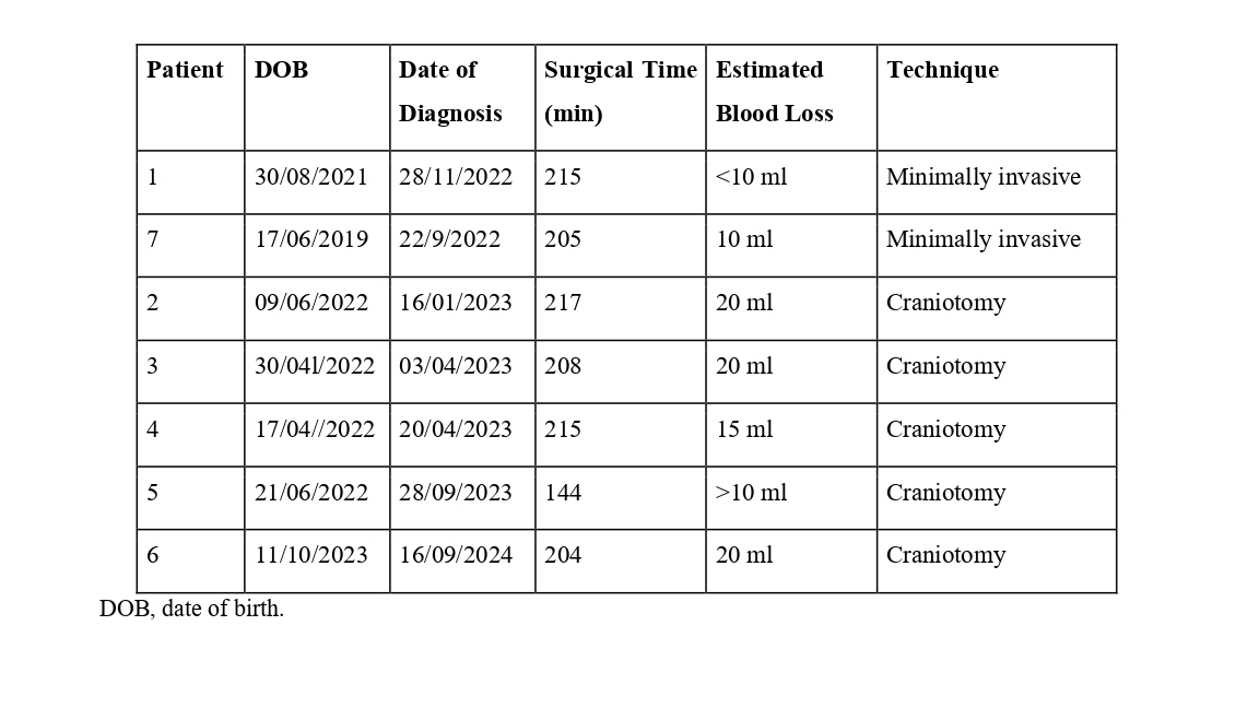
TABLE 1. Patient Demographic and Surgical Procedure Data.
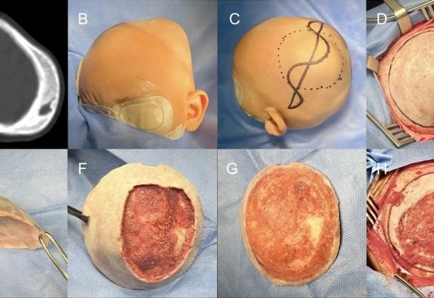
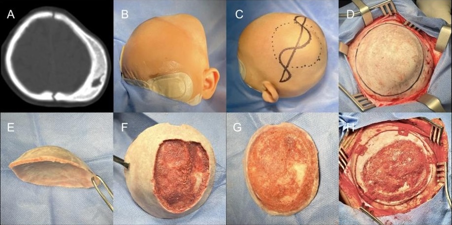
FIGURE 4. Image sequence of preoperative CT scan (A), intraoperative photos (B-G) and final surgical result before wound closure (H).
Postoperative photos were taken by a standardized manner. Parents were asked about their level of satisfaction regarding the esthetic result. In all cases, both parents and surgeons reported satisfactory results with no need of further procedures.
DISCUSSION
The management of cephalohematoma remains a subject of debate. While conservative management is the standard for small collections, our data suggest that early percutaneous aspiration (around week 3-4) is a safe and effective method to prevent the “egg-shell” calcification phase.
Although there is no consensus on what is defined as a small or large cephalohematoma, the authors decided to use the definition published by Üçer et al. (2021), which is the biggest observational cohort to our knowledge (94 patients) [2].
Xi et al. (2025) reported that the majority (83%) of cases required only a single puncture, with no cases of infection or ossification. Despite this data, parents are informed about the possibility of infection [1, 5] or repeated punctures if needed. Our data compares to international standards regarding infection rate.
Our described surgical technique for calcified lesions—performing a craniotomy for ex vivo burring—offers several advantages over simple in situ burring or flip over techniques. First, it allows for the assessment of bone thickness via transillumination, preventing accidental dural exposure. Second, it ensures a more symmetrical reconstruction of the cranial contour. Finally, delaying surgery until approximately 12 months of age, when the patient’s blood volume is higher, significantly reduces the risk of transfusion-related complications, as evidenced by our 0% transfusion rate.
Pediatric units should be encouraged to diagnose and refer promptly all patients with persistent cephalohematoma after two weeks of life, thus providing early treatment and avoidance of unnecessary open interventions and longer scars.
CONCLUSION
Management of cephalohematoma depends on the age of presentation and stage of calcification. A comprehensive algorithm is described for the entire spectrum of clinical presentation. Our ex vivo technique presents some advantages over previously published techniques and proves to be an effective and safe option for calcified lesions.
REFERENCES (5)
-
Ulma RM, Sacks G, Rodoni BM, et al. Management of calcified cephalohematoma of infancy: The University of Michigan 25-year experience. Plast Reconstr Surg. 2021;148(2):409-417. https://doi.org/10.1097/PRS.0000000000008199
-
Üçer M, Taçyıldız AE, Aydın I, et al. Observational case analysis of neonates with large cephalohematoma. Cureus. 2021;13(4):e14415. https://doi.org/10.7759/cureus.14415
-
Zimmermann P, Duppenthaler A. Infected cephalhaematoma in a five-week-old infant - case report and review of the literature. BMC Infect Dis. 2016;16(1):636. https://doi.org/10.1186/s12879-016-1982-4
-
Wong CH, Foo CL, Seow WT. Calcified cephalohematoma: Classification, indications for surgery and techniques. J Craniofac Surg. 2006;17(5):970-979. https://doi.org/10.1097/01.scs.0000229552.82081.de
-
Xi M, Shi H, Zhang G. Management of neonatal cephalohematoma and ossified cephalhematoma - 281 cases of personal 10-year experience. Childs Nerv Syst. 2025;41(1):77. https://doi.org/10.1007/s00381-024-06740-x