

Dermatogenic Necrotizing Fasciitis of the Neck: A Huge Skin Defect following Rare Etiology Purulent-Necrotic Phlegmon
Manuscript received 10 February 2026
Revised 28 March 2026
Accepted 09 April 2026
Available online 30 April 2026
J Diagn Treat Oral Maxillofac Pathol 2026;4: 100317
DOI: https://doi.org/10.23999/j.dtomp.2026.4.100317
Under a Creative Commons license
HOW TO CITE THIS ARTICLE
Rybak VA, Fesenko II. Dermatogenic necrotizing fasciitis of the neck: A huge skin defect following rare etiology purulent-necrotic phlegmon. J Diagn Treat Oral Maxillofac Pathol. 2026;10(4):100317.

To the Editor in Chief,
Necrotizing fasciitis (NF) is a rapidly progressive infection of fascial planes [1]. NF is also known as purulent-necrotic phlegmon in East-European states [2]. NF is an acute life-threatening infection, which causes necrosis of the subcutaneous tissue and the superficial fascial planes, leading to widespread gangrene and deep neck space purulent-necrotic process [3]. This process is often characterized by gas emphysema and crepitus (Aydin et al., 2025) [4] and can be termed as anaerobic phlegmon [2]. NF affects the head and neck in 1-10% of cases compared to the abdomen, groin, perineum, and extremities [5]. Gunaratne et al. (2018) in their systematic review and analysis of 1235 reported cases from the literature indicate that the etiology of cervical NF was odontogenic (47.04%), pharyngolaryngeal (28.34%), or tonsillar/peritonsillar (6.07%) [5]. According to Gupta et al. (2022), the source of infection upon NF of the neck was found to be odontogenic in 71.43% and in 28.57% it was due to peritonsillar abscess [1]. Other proven causes of cervical NF development were parotid infection, drug usage, hematogenous spread of infection, trauma, and iatrogenic reason [4, 6]. We would like to present a unique case of cervical NF from a small wound on the neck.
We present the case of a 73-year-old Caucasian woman was referred to our Center of Maxillofacial Surgery and Dentistry on September 05, 2011 with significant cervical edema, redness of the neck skin, and severe symptoms of intoxication. A wide area of blackened skin ran down the center of the front surface of the neck. According to the patient, it all started with a small wound on the front of her neck that she scratched while working in the field about a week ago. Crepitation was noted upon palpation of the inflamed area. Although some patients with NF have diabetes mellitus as a comorbidity, according to the history and laboratory data, this patient did not have diabetes mellitus. Against the background of preoperative drug detoxification preparation and antibacterial therapy under general anesthesia, the phlegmon was opened by evacuating a large volume of purulent exudate (approximately 45 ml) with gas bubbles through the removed area of blackened (necrotized) skin. Finger revision of the wound was also performed. During surgery, the tissues in the area of inflammation had an appearance typical of anaerobic phlegmon of soft tissues, where the muscles had the appearance of boiled meat with a large volume of necrotic masses. On the second day (Fig 1A), during dressing, a further partial sparing necrosectomy was performed, the wound was washed and ointment was applied. Minimally invasive debridement was performed daily until the wound was completely cleansed and granulations appeared (Fig 1B). Antibiotic therapy included ceftriaxone and metronidazole. Myramistin ointment was applied to the wound during dressings at the stages of cleansing the wound from purulent content and necrosis. From the third day after the operation, a decrease in hyperemia and swelling of the edges of the wound, as well as the area of the wound, was noted. The surface of the wound was deep in the middle.
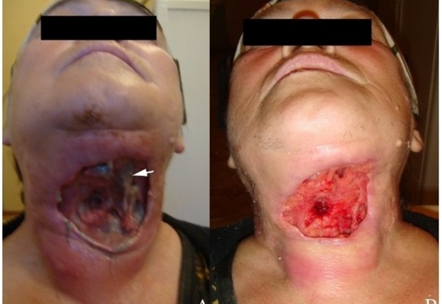
FIGURE 1. A 73-year-old female patient on the second (A) and fourth (B) days after going to the hospital and opening the purulent-necrotic phlegmon by removing the necrotic area of skin that had turned black, necrotic underlying tissues, and evacuating pus. Arrow shows necrotic tissues that were excised in stages sparingly with respect to local tissues. There is a significant skin defect.
Długosz-Karbowska et al. (2022) and Wagstaff et al. (2017) recommend using (1) a full- or split-thickness free flap, (2) a local flap, or (3) a biodegradable dermal substitute to reconstruct the lost soft tissues and skin on the neck [8, 9]. In our case, the patient was discharged with good general well-being and wound granulation for continued treatment in the plastic surgery department.
It is worth noting that NF is divided into two types according to pathogens: NF Type I - polymicrobial and NF Type II - Group A streptococcal (Vaid et al., 2002) [4]. In summary, we believe that the correct identification of the location of purulent content and its evacuation is key in the treatment of a similar pathology in the acute stage.
Declaration of Interests: The authors have no conflicts of interest to declare.
Informed Consent: Written consent was obtained from patient for publication of photographs.
Fundings: No funding was received for this study.
Peer-Review: Externally peer-reviewed.
AI Statement: In preparing this article, the authors used Google AI Mode to detail the process of gas bubbles formation in tissues and compare the terms necrotizing fasciitis and purulent-necrotic phlegmon for identity.
References (12)
-
Gupta V, Sidam S, Behera G, et al. Cervical necrotizing fasciitis: An institutional experience. Cureus. 2022;14(12):e32382. https://doi.org/10.7759/cureus.32382
-
Tymofieiev OO. Non-specific inflammatory diseases of soft tissues of the maxillofacial region. In: Tymofieiev OO, ed. Surgical dentistry and maxillofacial surgery. Vol 1. Marchenko TV; 2024:chap 5. Ukrainian.
-
Marginean E, Venter C, Domuta M, et al. Necrotizing fasciitis of the neck: Case presentation. J Mind Med Sci. 2017; 4(2):198-202. https://doi.org/10.22543/7674.42.P198202
-
Aydin A, Revend L, Revend D, et al. Necrotizing fasciitis of the head and neck - clinical features, diagnostics, and management strategies. Oncoscience. 2025;12:219-225. https://doi.org/10.18632/oncoscience.639
-
Gunaratne DA, Tseros EA, Hasan Z, et al. Cervical necrotizing fasciitis: Systematic review and analysis of 1235 reported cases from the literature. Head Neck. 2018;40(9):2094-2102. https://doi.org/10.1002/hed.25184
-
Dudde F, Kreibich M, Henkel KO. Cervicofacial necrotizing fasciitis of non-odontogenic origin. Dtsch Arztebl Int. 2024;121(5):147. https://doi.org/10.3238/arztebl.m2022.0407
-
Hohlweg-Majert B, Weyer N, Metzger MC, Schön R. Cervicofacial necrotizing fasciitis. Diabetes Res Clin Pract. 2006;72(2):206-208. https://doi.org/10.1016/j.diabres.2005.09.012
-
Długosz-Karbowska A, Smółka W, Markowski J. Cervical necrotizing fasciitis complicated by sepsis – case report. Pol Otorhino Rev. 2022;11(3):61-66. https://doi.org/10.5604/01.3001.0016.0174
-
Wagstaff MJ, Caplash Y, Greenwood JE. Reconstruction of an anterior cervical necrotizing fasciitis defect using a biodegradable polyurethane dermal substitute. Eplasty. 2017;17:e3.
-
Rajmohan S, Gao C, Rajmohan K, et al. A parotid abscess out of control resulting in craniocervical necrotising fasciitis in the context of diabetes mellitus-a case report and review of the literature. Gland Surg. 2024;13:257-264. https://doi.org/10.21037/gs-23-365
-
Abdurrazaq TO, Ibikunle AA, Braimah RO. Cervical necrotizing fasciitis: A potentially fatal disease with varied etiology. Ann Med Health Sci Res. 2016;6(4):251-256. https://doi.org/10.4103/amhsr.amhsr_33_16
-
Vaid N, Kothadiya A, Patki S, Kanhere H. Necrotising fasciitis of the neck. Indian J Otolaryngol Head Neck Surg. 2002;54(2):143-145. https://doi.org/10.1007/bf02968735