
March 31, 2020
https://doi.org/10.23999/j.dtomp.2020.3.3
J Diagn Treat Oral Maxillofac Pathol 2020;4:60−1.
Under a Creative Commons license
HOW TO CITE THIS ARTICLE
Beridze B, Cherniak OS. Large mucocele in the labial and buccal mucosa. J Diagn Treat Oral Maxillofac Pathol 2020;4(3):60–1.
ARTICLE
PANEL A.
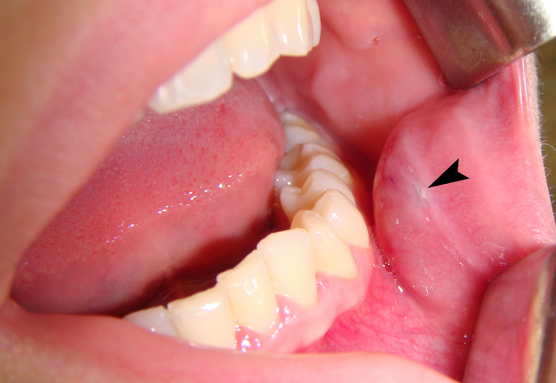
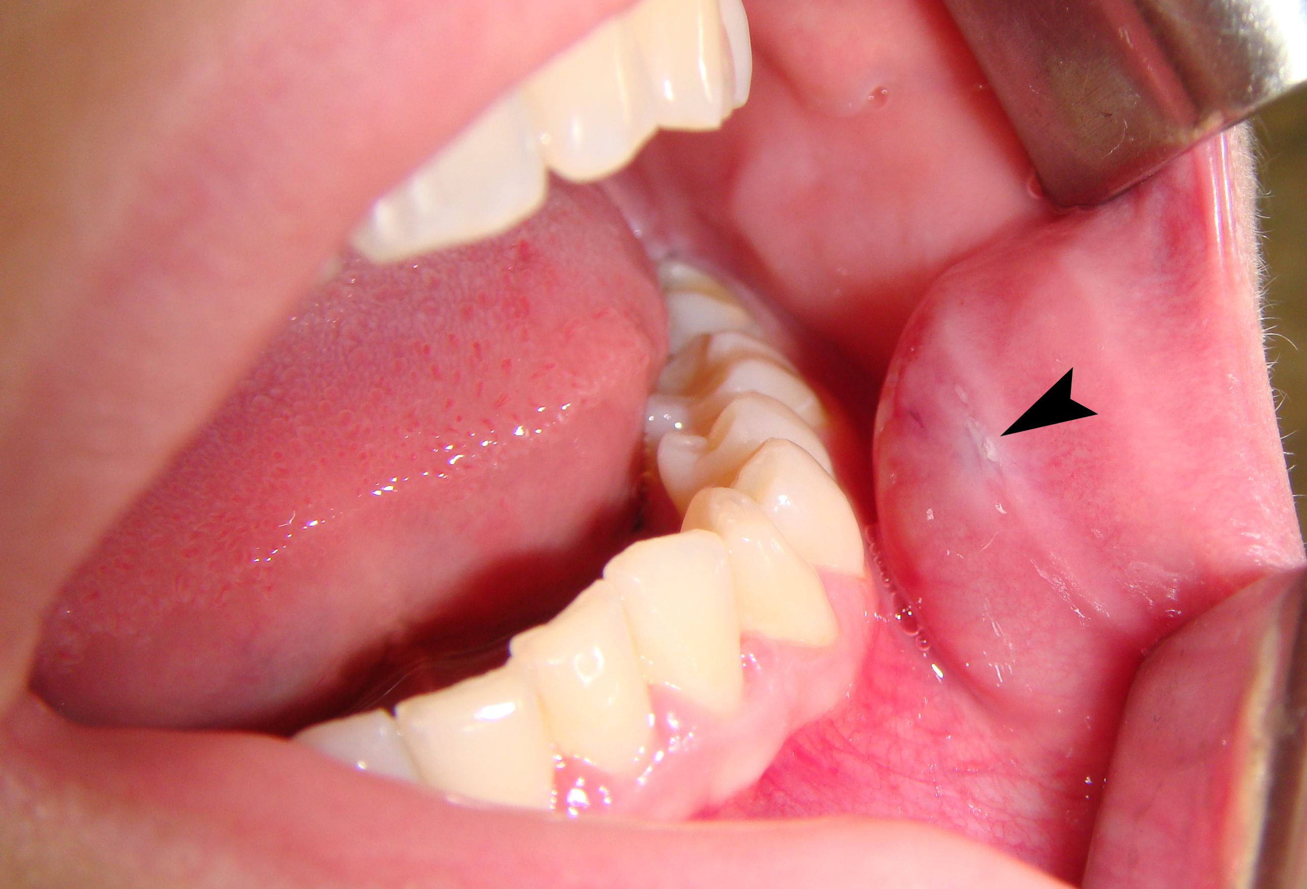
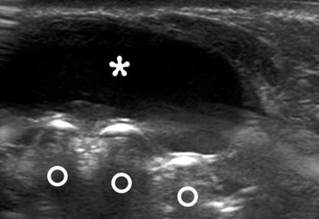
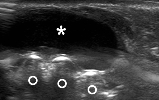
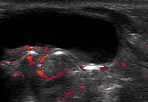
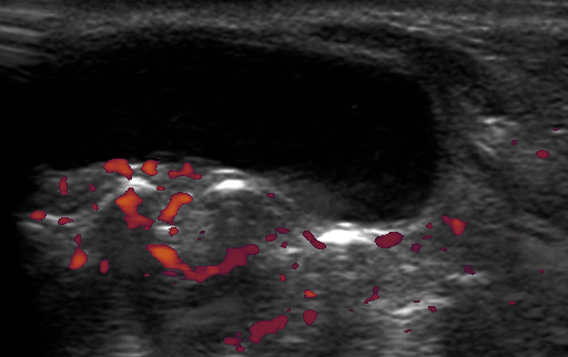
A 26-year-old woman presented to the maxillofacial surgery clinic with painless oval shape lesion (Panel A, arrow) arisen from the lower lip and extended to the buccal mucosa. According to the patient, the mass arose after repeated trauma (biting) of the mucosa (Panel B, arrowhead) approximately 3 months ago. Once the patient noted that when she bit the mass, it`s emptied, but then began to grow again. Upon intraoral examination the lesion measured 1 × 1.8 cm in size. Palpation showed its soft and spongy texture. No bluish pattern of the surface was noted. B-mode (Panel C) and power Doppler (Panel D) ultrasound demonstrated cystic, avascular lesion measuring 1.71 × 0.92 cm with echogenic content (asterisk), no signs of echogenic debris, and distinct margins. Acoustic shadowing behind the hyperechogenic vestibular surface of the lower teeth is labeled by circles. The depth of the cropped sonograms is 1.56 cm.
PANEL B.
PANEL C.
PANEL D.
Mucocele of the oral cavity (synonyms: oral mucocele, mucous cyst, retention cyst, retention cyst of the minor salivary gland, mucous retention cyst) is a cystic lesion of the minor salivary gland due to its duct alternation/inflammation and subsequent accumulation of saliva. Differential diagnostics of mucocele is usually performed with other similar oral masses: lipomas, lymphangiomas, and hemangiomas. Removal of mucocele includes the excision of mucous cyst associated overlying mucosa, own glandular tissue and other minor salivary glands which are visualized in the wound. Histopathologically, two types of oral mucocele are distinguished: retention and the more often, extravasation variant. Summarizing, despite the majority of mucocele cases presented with mucosa surface color ranged from deep blue to light blue, our case clearly shows a mucocele with a pink color of mucosa above. Recurrence is a complication usually associated with a violation of the operation technique. ■ DTJournal.org