Digitalization: Replacement of Upper Lateral Incisor
Published September 25, 2025
J Diagn Treat Oral Maxillofac Pathol 2025; 9: 100309.
DOI: 10.23999/j.dtomp.2025.9.100309
Under a Creative Commons license (CC BY-NC-SA 4.0)
HOW TO CITE THIS ARTICLE
Mastakov OY, Shkilniak N. Digitalization: Replacement of upper lateral incisor. J Diagn Treat Oral Maxillofac Pathol. 2025 Sep;9(9):100309. https://doi.org/10.23999/j.dtomp.2025.9.100309

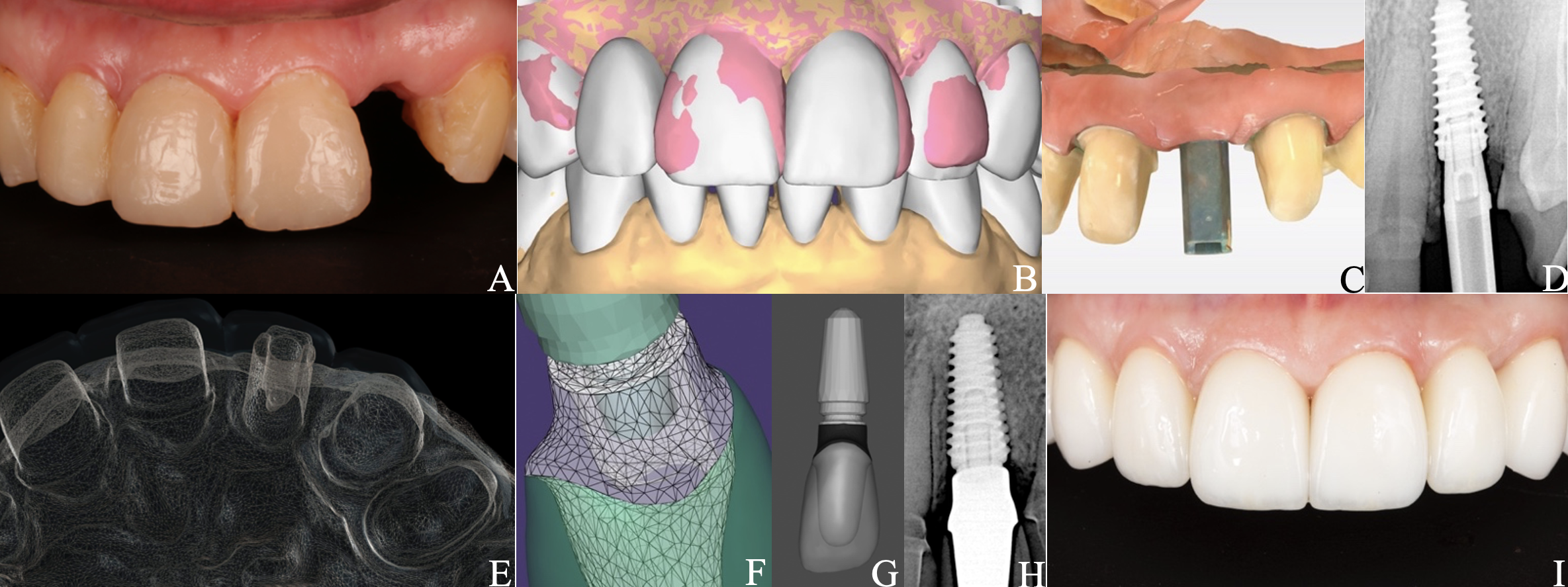
PANELS A-I.
A 50-year-old woman with a fracture of the tooth 2.2 (Pan A) was referred to our clinic. After cone-beam computed tomography, atraumatic root extraction with preservation of the vestibular bone wall was performed. A sequential drilling protocol using the AnyRidge® system (MegaGen) along the palatal wall of the socket was applied for an implant size of 4.0×11.5 mm. Upon the implantation navigation was not used. Additionally, bone grafting of the socket was performed with a xenograft (BMG-20, BIO-GEN®; BIOTECK). The next step was to use a free gingival graft (FGG) harvested from right maxillary tuberosity (RMT) for the “poncho” technique. At the time of implant opening, a preliminary scan (CEREC® Primescan AC; Dentsply Sirona) for a digital wax-up of the tooth anatomy (Pan B, DentalCAD 3.1 Rijeka [exocad]) considering the patient's aesthetic preferences was performed. Implant opening was done using the roll technique and installing an individual modified healing abutment (composite [Gradia® Direct AO3; GC] on a titanium platform, made clinically). The repeated gingival plasty, FGG from the RMT, was also performed. Panels C and D show control scan of the maxillary teeth with scan marker on the implant and digital radiography (Genoray X-PORT-IV-e; Genoray Co) four weeks before permanent prosthetics. Panel E illustrates evaluation and verification of the project in Smilecloud (Smilecloud SRL). A platform marker from a Zero Bone Loss Concepts Kit® (MegaGen) was used to select the appropriate height of the permanent platform (ZrGEN 4015, Ø4.0, AnyOne®; MegaGen) on the individual zirconia abutment (Pan F, DentalCAD and exocad webview 1.6.18). On top of which an InitialTM LiSi Press (GC) lithium disilicate crown (Pan G, DentalCAD) was cemented in the laboratory after fitting for optimal aesthetics. Screw fixation of the restoration to the implant was done at 35 Ncm torque. An adhesive protocol was applied for lithium disilicate ceramic restorations on the teeth. Panel H highlights control radiography at month six. Intraoral condition after 6 months (Pan I) showed nice pink-white aesthetics. Panels A and I were obtained using Canon EOS 6D full-frame digital camera (Canon) with Canon Macro Twin Lite MT-26-EX-RT.