Can Articles from the Images Section Be Cited in Journals Indexed in Scopus? Launch of a New Images Section Focused on Digitalization
Published September 30, 2025
J Diagn Treat Oral Maxillofac Pathol 2025; 9: 100310.
DOI: 10.23999/j.dtomp.2025.9.100310
Under a Creative Commons license (CC BY-NC-SA 4.0)
HOW TO CITE THIS ARTICLE
Tymofieiev OO, Fesenko II, Fil V. Can articles from the Images section be cited in journals indexed in Scopus? Launch of a new Images section focused on digitalization. J Diagn Treat Oral Maxillofac Pathol. 2025 Sep;8(9):100310. https://doi.org/10.23999/j.dtomp.2025.9.100310

INSTITUTIONAL REPOSITORY
https://ir.kmu.edu.ua/handle/123456789/830
SUMMARY
In this editorial, we analyze articles of the “Images” type in other peer-reviewed journals as well as the 6-year experience of publishing such articles in the Journal of Diagnostics and Treatment of Oral and Maxillofacial Pathology (JDTOMP). We also describe the experience of citing articles from the JDTOMP and The New England Journal of Medicine (NEJM) in the Scopus database. This is important for every peer-reviewed publication because one of the notes and conditions for indexing a journal in Scopus is citations of its articles in good journals indexed in Scopus. Taking into account the experience of journal in publishing 19 articles of the Images type, it was decided to expand the direction of publishing short manuscripts, but with a slightly different focus. A new section called “Images: Digital” has been launched, which aims to publish the same ultra-short articles, but not dedicated to pathology, but specifically dedicated to digital solutions in oral and maxillofacial surgery.
KEY WORDS
Article type, Images, Scopus, database, digital, section, editor, digitalization
EDITORIAL
Ultra-short articles in sections “Images” or “Pictures” are a popular type of articles in various medical peer-reviewed journals [1-5]. This is usually a one-page article without a reference list [5], but sometimes the article can be 2 or 3 pages long [1, 6] and have references [4]. However, in our humble opinion, the gold standard for publishing articles of this type has been achieved by The New England Journal of Medicine (NEJM) [5, 7]. In their journal, this type of article is called “Images in Clinical Medicine.” The purpose of this type of article is to simplify the preparation and reduce the time frame for writing the manuscript, while highlighting a rare pathology and presenting a treatment option. Another goal is to provide readers with a quick introduction to the clinical case. Inspired by the example of the NEJM, in 2019 a similar section titled "Pictures in Oral and Maxillofacial Surgery" was also launched in the Journal of Diagnostics and Treatment of Oral and Maxillofacial Pathology (JDTOMP) [8]. And during the period from July 1, 2019, to September 1, 2025, that is, for more than 6 years, 19 Images articles were published in the JDTOMP [6, 9-26]. Out of 19 articles, 2 articles have videos [16, 22]. Most of the manuscripts in this section were published by authors from Ukraine. One article from Qatar and another joint article by authors from Ukraine and Georgia [6, 21].
We know that the success of this section would not have been possible without the confident leadership of the editor of this section, Dr. Camilo Mosquera from the University of Texas Medical Branch at Galveston [8].
Publishing a concise and meaningful article is of great importance to clinicians. However, the editorial board of each peer-reviewed journal must understand whether a given article can bring dividends to the journal in the form of citations. Moreover, one of the notes and conditions for indexing a journal in Scopus is citations of its articles in good journals indexed in Scopus (email response according to title evaluation process from Scopus Title Evaluation Team, July 2022).
The experience of the NEJM proves that articles of the “Images” type are successfully cited in journals indexed by Scopus [7, 27]. In particular, an article entitled “Lemierre's Syndrome” (Walkty and Embil, 2019) has received 23 citations (Fig 1) in other journals indexed by Scopus since its publication (i.e., 6 years ago). Articles of type “Images” of the JDTOMP also demonstrate the presence of citations in journals indexed by Scopus (Figs 2 and 3) [16, 28]. Although of course the frequency of citations is also affected by the availability of journal articles for search by authors in PubMed/PubMed Central according to the journal sample. Although, of course, the frequency of citations is also affected by the availability of journal articles for search by authors in PubMed/PubMed Central, as is the case with the NEJM [29].
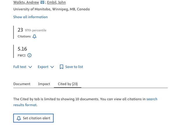
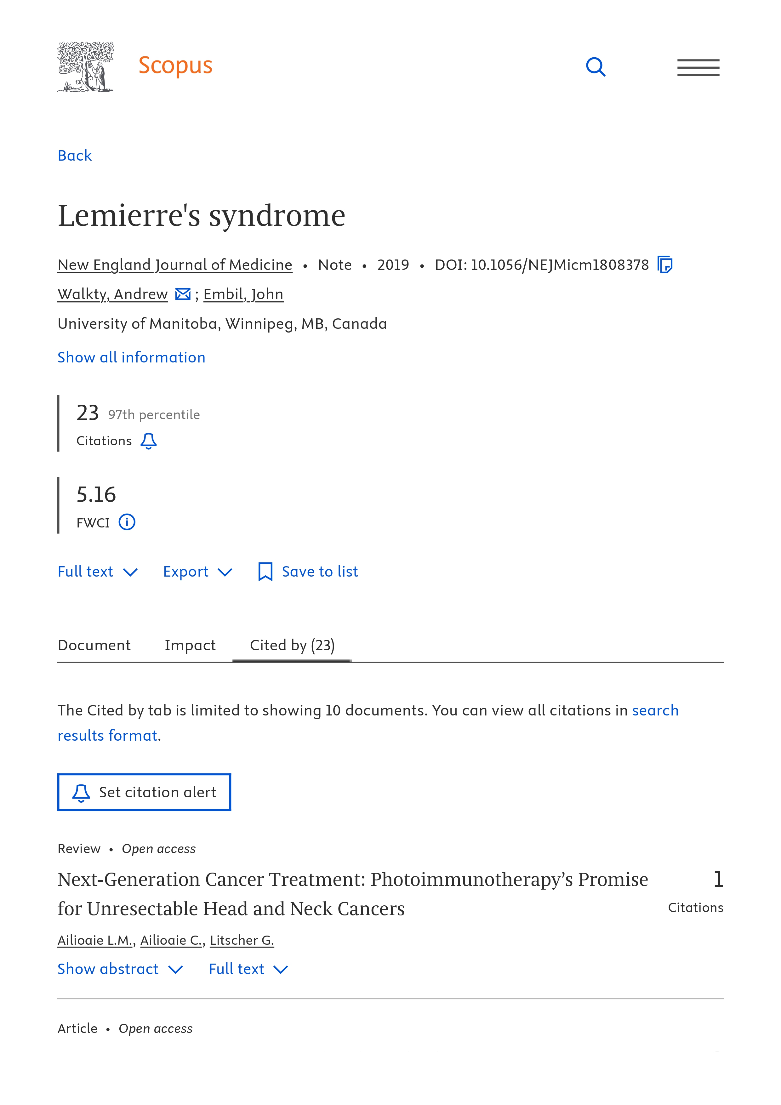
FIGURE 1. An example of numerous citations in the Scopus database of an article from the section “Images in Clinical Medicine” of The New England Journal of Medicine [7, 27].
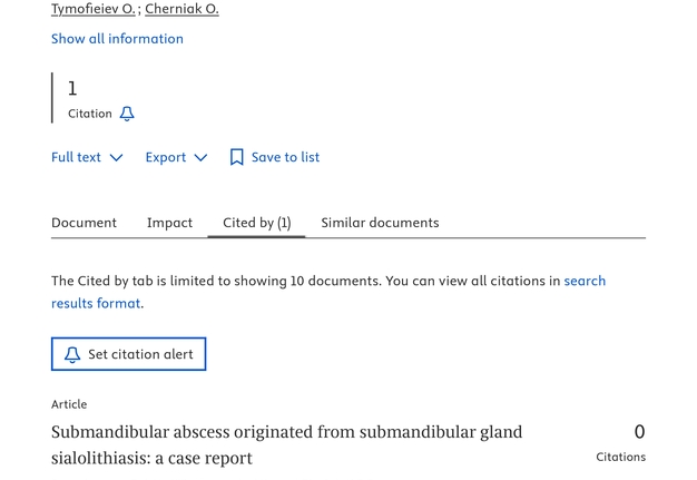
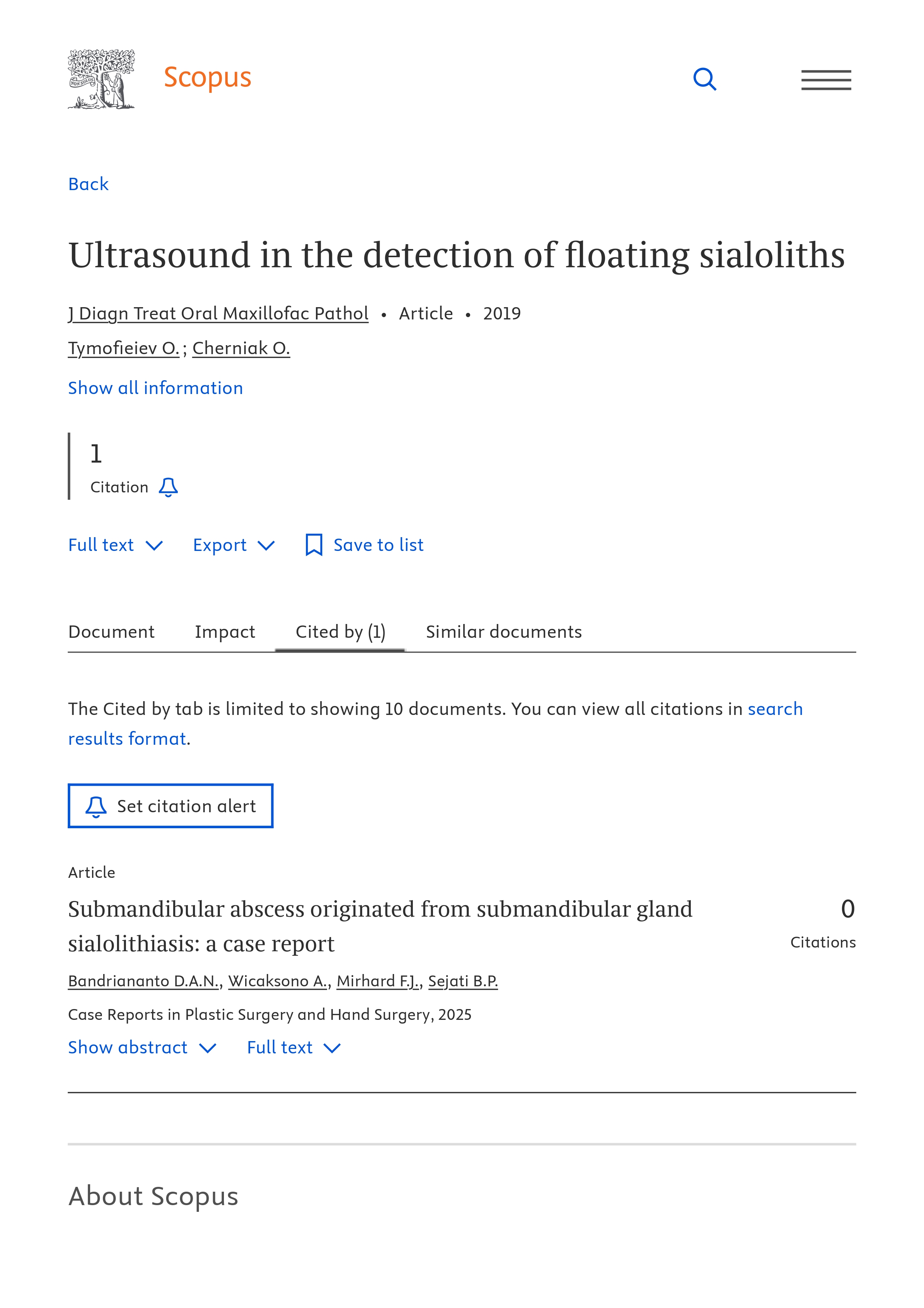
FIGURE 2. An example of citation in the Scopus database of an article from the section “Images” of the Journal of Diagnostics and Treatment of Oral and Maxillofacial Pathology [16, 28].
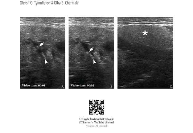
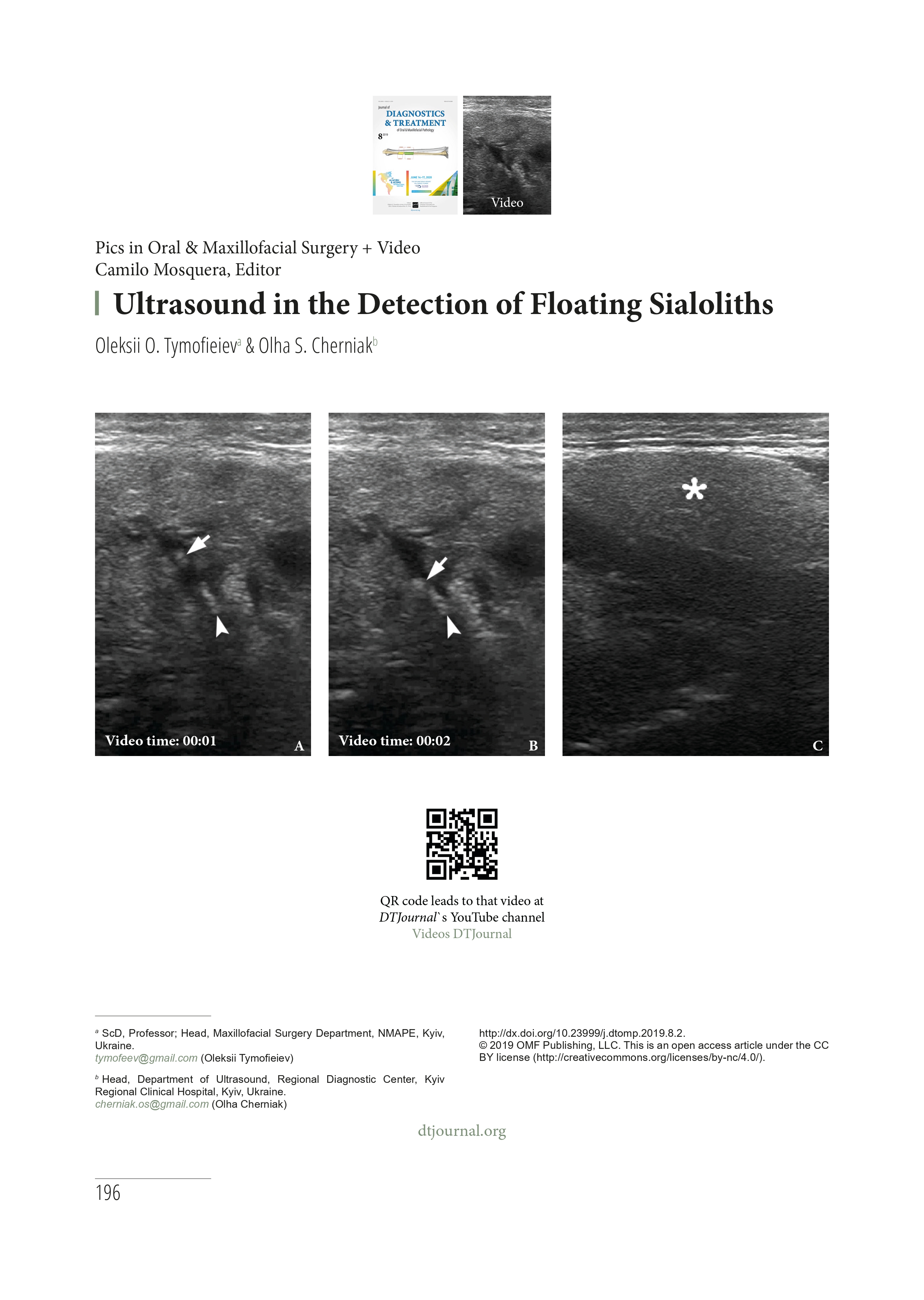
FIGURE 3A. An example of what a cited 2-page article (A, B) of type “Images” in the Journal of Diagnostics and Treatment of Oral and Maxillofacial Pathology (Tymofieiev and Cherniak, 2019) looks like [16].
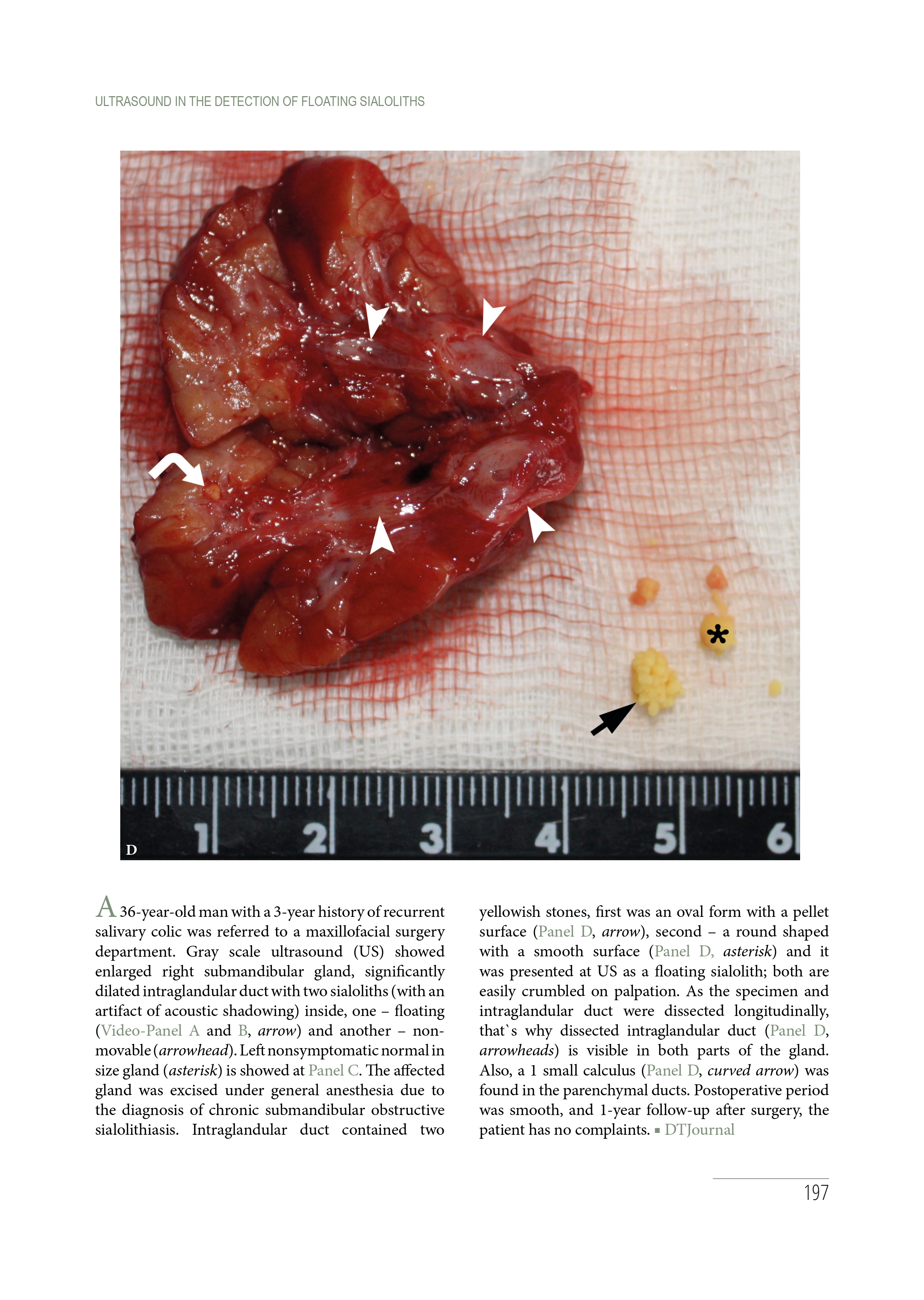
FIGURE 3B. An example of what a cited 2-page article (A, B) of type “Images” in the Journal of Diagnostics and Treatment of Oral and Maxillofacial Pathology (Tymofieiev and Cherniak, 2019) looks like [16].
Summarizing the above, it would be correct to say that articles of the “Images” type can and do definitely get cited [7, 16, 27, 28]. This fact is the basis for our Journal to introduce a new section with a similar type of articles, but which focus on digital solutions. Digitalization at all stages of diagnosis, planning and treatment in dentistry, oral and maxillofacial surgery is becoming increasingly widespread [29-31]. The range of applications of digital solutions is extremely wide, including digital workflow and tooth replacement methods [31]. And even, digital workflow and jaw in a day microvascular technique [32]. That's why, starting in September 2025, we are accepting manuscripts in this new required section titled “Images: Digital” [33]. The section editor was chosen by a specialist who regularly implements digital technologies in clinical practice and has experience in publications, namely Dr. Oleg Mastakov from Kyiv, Ukraine.
Thus, our editorial team is happy to begin a new, next stage of the Journal’s development by adding a new, much-needed section. We invite everyone to submit unique manuscripts for peer-review. Our team will be happy to provide the highest standards of review process.
There is no alternative to digital transformation.
—Jeff Bezos
Founder of the Amazon, Inc.
REFERENCES (33)
-
Cortés Arciniegas A, Almanzo S, et al. Multiple submandibular gland sialolithiasis: An unusual presentation of a common condition. Med Clín Práct. 2025;8(3):100502. https://doi.org/10.1016/j.mcpsp.2025.100502
-
Sanfilippo CJ, Javaheri M, Handler S, et al. Benign lobular inner nuclear layer proliferations of the retina associated with congenital hypertrophy of the retinal pigment epithelium. Ophthalmology. 2023;130(3):265-273. https://doi.org/10.1016/j.ophtha.2022.10.011
-
Chen G, Peng P. Cinematic rendering of Klippel-Trénaunay syndrome. Radiology. 2025;316(2):e250230. https://doi.org/10.1148/radiol.250230
-
Senapati A, Saleh Y, Chang SM. Patent foramen ovale and dysarthria in a man in his 60s. JAMA Cardiol. 2025;10(9):961. https://doi.org/10.1001/jamacardio.2025.1679
-
Karkhur S, Ayush Gupta A. Ocular gnathostomiasis. N Engl J Med. 2025;393(7):e9.https://doi.org/10.1056/NEJMicm2502742
-
Grimaldi Finol GA, Hawamdeh S, Al Khalil M. Extensive and advanced craniofacial dysplasia. J Diagn Treat Oral Maxillofac Pathol 2022;6(12):158-160. https://doi.org/10.23999/j.dtomp.2022.12.2
-
Walkty A, Embil J. Lemierre's Syndrome. N Engl J Med. 2019;380(12):e16. https://doi.org/10.1056/nejmicm1808378
-
Tymofieiev OO, Fernandes RP. The New England Journal of Medicine: Images in clinical medicine: A role model section for DTJournal. J Diagn Treat Oral Maxillofac Pathol 2019;3(7):A11. https://doi.org/10.23999/j.dtomp.2019.7.1
-
Demidov VH, Cherniak OS. Hemolymphangioma of the neck. J Diagn Treat Oral Maxillofac Pathol 2022;6(8):114-116. https://doi.org/10.23999/j.dtomp.2022.8.2
-
Rybak VA. Severe self-inflicted gunshot wound of the face. J Diagn Treat Oral Maxillofac Pathol 2022;6(4):63-64. https://doi.org/10.23999/j.dtomp.2022.4.2
-
Shamova TO, Blyzniuk VP. Gunshot fracture of the mandible. J Diagn Treat Oral Maxillofac Pathol 2022;6(4):65-66. https://doi.org/10.23999/j.dtomp.2022.4.3
-
Fesenko II. Rubber bullet-induced wound of the cheek. J Diagn Treat Oral Maxillofac Pathol 2022;6(4):67-68. https://doi.org/10.23999/j.dtomp.2022.4.4
-
Cherniak OS, Fesenko II. Periorbital abscessing furuncle. J Diagn Treat Oral Maxillofac Pathol 2021;5(1):13-14. https://doi.org/10.23999/j.dtomp.2021.1.4
-
Shamova TO. Unilateral ‘Sausaging’ of the Stensen's duct. J Diagn Treat Oral Maxillofac Pathol 2019;3(8):201. https://doi.org/10.23999/j.dtomp.2019.8.5
-
Nagorniak IV. Lateral sinus lift. J Diagn Treat Oral Maxillofac Pathol 2020;4(9):178. https://doi.org/10.23999/j.dtomp.2020.9.4
-
Tymofieiev OO, Cherniak OS. Ultrasound in the detection of floating sialoliths. J Diagn Treat Oral Maxillofac Pathol 2019;3(8):196-197. https://doi.org/10.23999/j.dtomp.2019.8.2
-
Cherniak OS, Nozhenko OA. Understanding the head and neck ultrasound: From simple to complicated cases: Submandibular abscess. J Diagn Treat Oral Maxillofac Pathol 2020;4(8):198-199. https://doi.org/10.23999/j.dtomp.2019.8.3
-
Nagorniak IV. Clinical appearance of lateral incisive canal. J Diagn Treat Oral Maxillofac Pathol 2019;3(8):200. https://doi.org/10.23999/j.dtomp.2019.8.4
-
Shamova TO, Pavlenko RA. Post-traumatic facial and intracranial emphysema. J Diagn Treat Oral Maxillofac Pathol 2020;4(5):95–6. https://doi.org/10.23999/j.dtomp.2020.5.2
-
Demidov VH, Rybak VA. Giant parotid pleomorphic adenoma. J Diagn Treat Oral Maxillofac Pathol 2020;4(3):62-63. https://doi.org/10.23999/j.dtomp.2020.3.4
-
Beridze B, Cherniak OS. Large mucocele in the labial and buccal mucosa. J Diagn Treat Oral Maxillofac Pathol 2020;4(3):60-61. https://doi.org/10.23999/j.dtomp.2020.3.3
-
Demidov VH, Ripolovska OV. How multiple the submandubular gland sialoliths can be? J Diagn Treat Oral Maxillofac Pathol 2019;3(7):174-175. https://doi.org/10.23999/j.dtomp.2019.7.2
-
Fesenko II. Abscess of the left tongue. J Diagn Treat Oral Maxillofac Pathol 2020;4(2):39-40. https://doi.org/10.23999/j.dtomp.2020.2.5
-
Khadem AA. Infected nasolabial cyst. J Diagn Treat Oral Maxillofac Pathol 2020;4(2):38. https://doi.org/10.23999/j.dtomp.2020.2.4
-
Fedorenko PA, Shamova TO. Eagle syndrome: Symptomatic elongated styloid process. J Diagn Treat Oral Maxillofac Pathol 2020;4(1):21. https://doi.org/10.23999/j.dtomp.2020.1.3
-
Ivanchenko OO, Demidov VH. Parotid gland lipoma in a 52-year-old patient. J Diagn Treat Oral Maxillofac Pathol 2020;4(1):22. https://doi.org/10.23999/j.dtomp.2020.1.4
-
Scopus: Lemierre's Syndrome [internet]. 2019 [cited 2025 Sep 24]. Available from: https://www.scopus.com/pages/publications/85063327344?origin=resultslist#tab=citedBy
-
Scopus: Ultrasound in the detection of floating sialoliths [internet]. 2019 [cited 2025 Sep 25]. Available from: https://www.scopus.com/pages/publications/105001986612?origin=resultslist#tab=citedBy
-
PubMed: Lemierre's Syndrome [internet]. 2019 [cited 2025 Sep 24]. Available from: https://pubmed.ncbi.nlm.nih.gov/30893539/
-
Mykhaylyuk N. Digitalization: New era of dentistry. J Prosthet Dent. 2024 Jun;131(6):988-989. https://doi.org/10.1016/j.prosdent.2024.03.002
-
Garcia-Torres F, Jurado CA, Rojas-Rueda S, et al. Immediate implant therapy with full-gigital workflow to replace a central incisor. Dent J. 2025;13(2):73. https://doi.org/10.3390/dj13020073
-
Mosquera C, Marwan H. Jaw in a Day: How to perform your first case—our workflow. Craniomaxillofac Trauma Reconstr. 2025;18(3):38. https://doi.org/10.3390/cmtr18030038
-
Mastakov OY, Shkilniak N. Digitalization: Replacement of upper lateral incisor. J Diagn Treat Oral Maxillofac Pathol. 2025;9(9):100309. https://doi.org/10.23999/j.dtomp.2025.9.100309