

Recurrent Sialolithiasis following Submandibular Gland Excision: Ultrasonography
Manuscript received 14 February 2026
Revised 07 April 2026
Accepted 16 April 2026
Available online 31 May 2026
J Diagn Treat Oral Maxillofac Pathol 2026;5: 100318
DOI: https://doi.org/10.23999/j.dtomp.2026.5.100318
Under a Creative Commons license
HOW TO CITE THIS ARTICLE
Mastakov OY, Fesenko II, Cherniak OS. Recurrent sialolithiasis following submandibular gland excision: Ultrasonography. J Diagn Treat Oral Maxillofac Pathol. 2026;10(5):100318.

ABSTRACT
In this report, we present a 69-year-old Caucasian female patient with recurrent sialolithiasis following submandibular gland excision (six years ago). Ultrasonography showed and enlarged Wharton’s duct measured 2.17 (length) × 0.67 (diameter) cm. A 5 × 3-mm sialodocholith was removed upon ductotomy with duct remnant preservation. In the analyzed and available literature covering sialodochitis of the residual Wharton’s duct and recurrence of sialolithiasis after sialoadenectomy, we did not find a single descriptive ultrasound case with a presented sonogram. Thus, to our knowledge this is the first case with ultrasound presentation and description of the condition of the tissues of the floor of mouth in a patient with such recurrence. The article also discusses the surgical tactics of the international surgical community in the treatment of similar cases.
KEY WORDS
Sialolithiasis, submandibular gland, Wharton’s duct, sialoadenectomy, ultrasonography, sialodochitis, sialodocholith
INTRODUCTION
The formation of calculi in the salivary glands and their ducts, i.e. sialolithiasis, is the most common disease of the major salivary glands [1]. Calculous submaxillitis is observed in 98.2% of patients with calculous sialadenitis [2]. Recently, the rate of submandibular gland removal due to sialolithiasis has significantly decreased to below 5% [3]. However, the following early and late complications may occur after sialoadenectomy. Such as infection, bleeding, residual inflammation in Wharton’s duct, unesthetic scar, neurological complications, etc. [4]. Tymofieiev (2025) notes that in the early postoperative period after removal of submandibular glands, in some cases, patients from other institutions with inflammatory manifestations in the postoperative wound area were referred [2]. Radiological examination revealed unremoved calculi, which required repeated surgical treatment. Berini-Aytes and Gay-Escoda (1992) analyzed postoperative complications in 206 submandibular gland excisions (62% due to sialolithiasis) and found residual inflammation in Wharton’s duct (i.e., sialodochitis) in 7.3% (i.e., 15 glands) of cases [4].
Several cases of recurrent sialolithiasis in the residual part of the Wharton’s duct have already been described [3, 5-11], but the literature does not present ultrasound data in such cases. The purpose of this paper is to highlight the clinical and first-time ultrasonographic data in surgical treatment of recurrent sialolithiasis following submandibular gland excision.
CASE REPORT
A 69-year-old Caucasian woman with history of chronic sialadenitis and right submandibular gland sialolithiasis six years status-post gland excision (Fig 1) presented with swelling posterior and lateral to the right sublingual fold (Fig 2) and floor of mouth pain. Saliva secretion from the orifice of the right submandibular gland duct was not noted. Salivary stone was not palpated in the oral cavity.
When referring the hospital on November 03, 2014, a gray scale and color Doppler ultrasonography (HD11 XE; the Philips) was performed. According to longitudinal ultrasound the proximal aspect of the right remnant Wharton’s duct showed a dilation up to a maximum of 0.67 cm (Fig 3). The total length of the dilated duct was 2.17 cm. Transverse ultrasound of the duct showed it to be round with dimensions of approximately 0.7 × 0.7 cm. This massive Wharton’s duct dilatation corresponded to signs of sialodochitis (also known as ductocele [2]). No ultrasound evidence of a stone was found. According to ultrasound imaging, the right submandibular gland was absent, and the left was unchanged. Two reactive lymph nodes (slightly enlarged) were noted in the right submandibular region (Fig 3B). Due to the lack of reliable ultrasound data on the presence of a sialolith in the remnant duct of the submandibular gland or in the sublingual gland, it was decided to prescribe anti-inflammatory therapy and monitor the dynamics by observing the patient in the hospital.
On the third day after hospitalization (i.e., on November 06, 2014), a decision was made to perform a ductotomy due to the increase in symptoms of inflammation around right sublingual fold, namely the increase in edema and pain. Applying ductotomy under the local anesthesia, a gray-transparent liquid content was obtained and an elongated sialodocholith measuring 5 × 3 mm was removed (Fig 4). In the days following calculus removal, a gradual decrease in swelling and complaints was noted.
Figure 5 compares gray scale sonograms (HD11; the Philips) of the right and left submandibular areas on the day 6 (November 09, 2014) from the moment of the patient’s hospitalization and on the day 3 (November 06, 2014) from the moment of the calculus removal. The absence of the right submandibular gland and the presence of an unchanged left submandibular gland were evidenced.
One month after the intervention, the patient had no complaints.
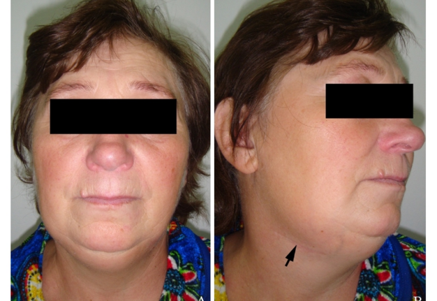
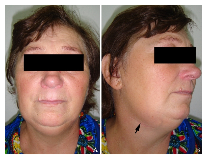
FIGURE 1. (A) Anterior view of a 69-year-old Caucasian female with recurrent sialolithiasis following submandibular gland excision. Photos when going to the hospital as of November 03, 2014. The skin scar from sialoadenectomy performed 6 years ago is indicated by an arrow (B).
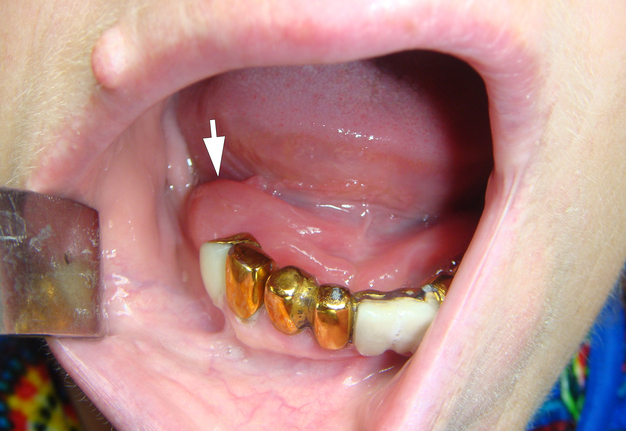
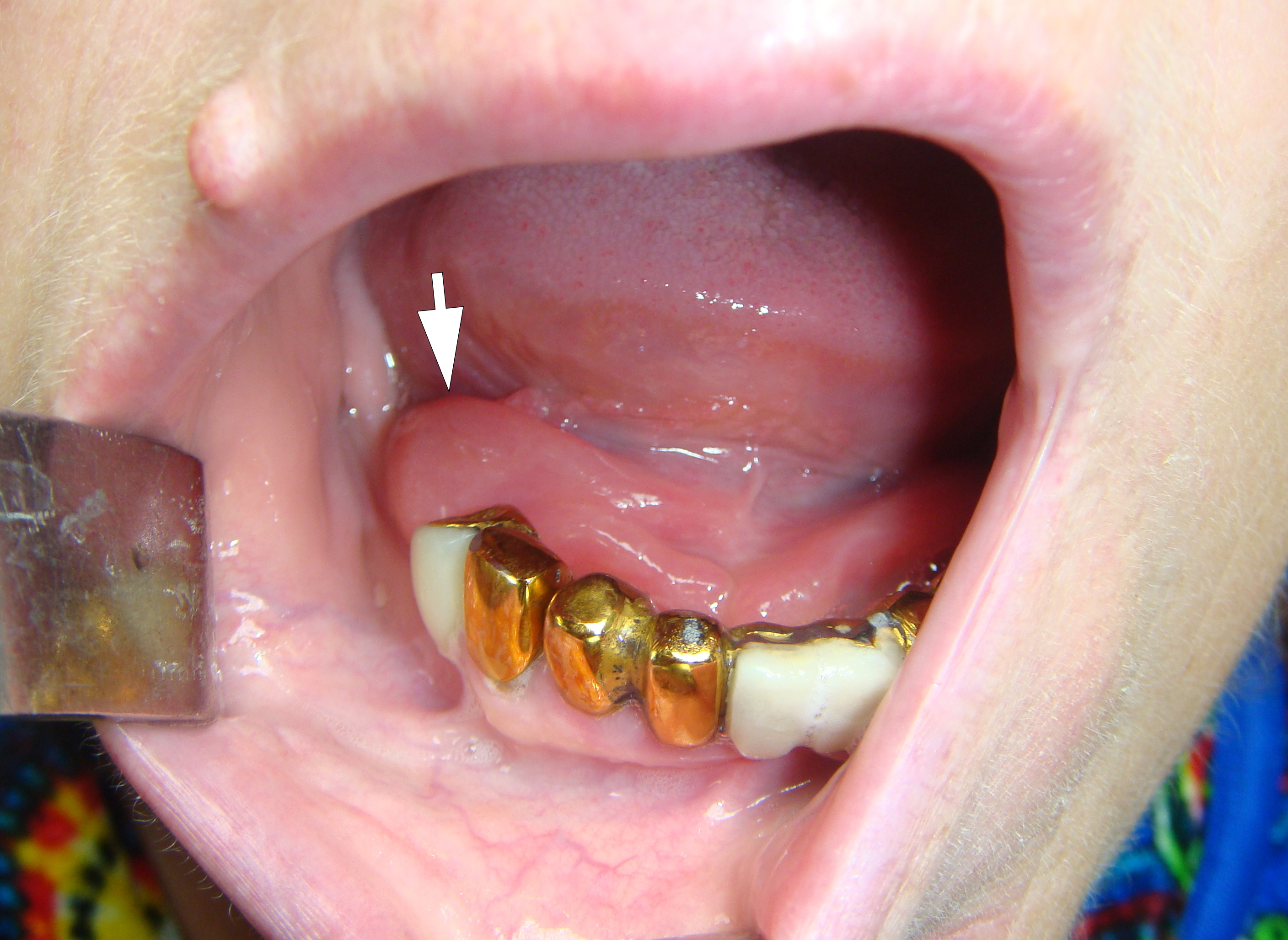
FIGURE 2. Intraoral view shows swelling (arrow) posterior and lateral to the right sublingual fold.
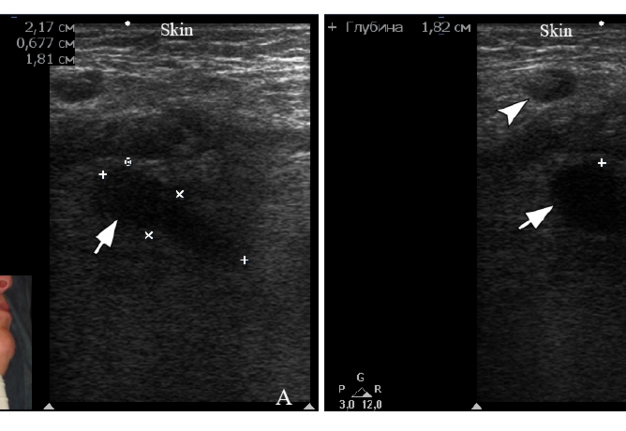
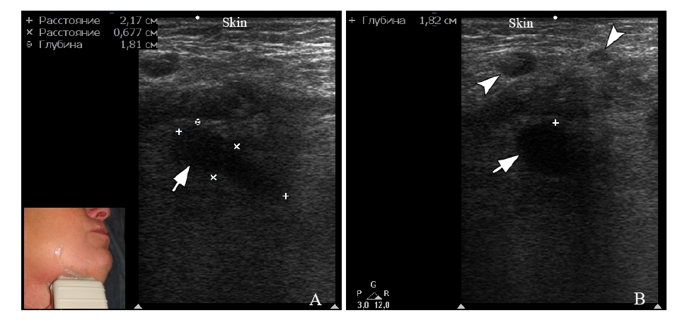
FIGURE 3. (A) The longitudinal gray scale ultrasound of the massively dilated right Wharton’s duct (arrow) measured 2.17 × 0.67 cm. The length of the duct is indicated by “+” calipers and width is indicated by “×” calipers. Ultrasound appearance consistent with sialodochitis (also known as ductocele). (B) The transversal gray scale ultrasound shows a cross-section of the duct (arrow). Arrowheads label two reactive submandibular lymph nodes. The depth of the expanded duct from the skin surface is 1.82 cm and is marked with a “+” caliper. The “depth” of ultrasonography is 5.0 cm.
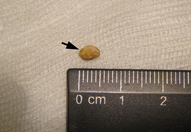
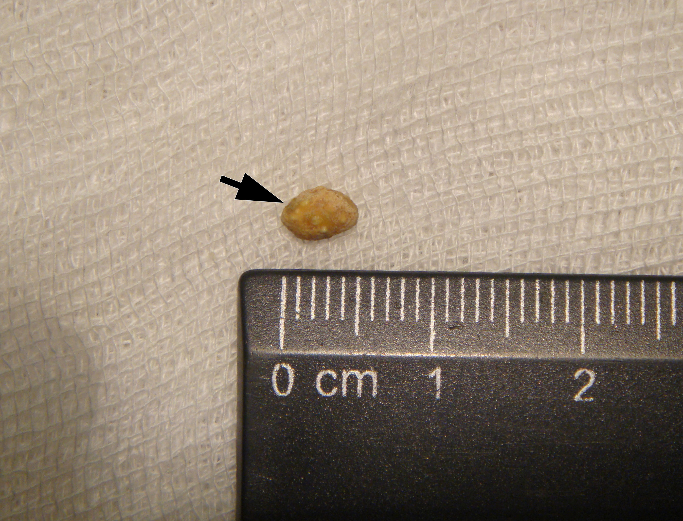
FIGURE 4. A 5 × 3-mm sialodocholith (arrow) removed during ductotomy.
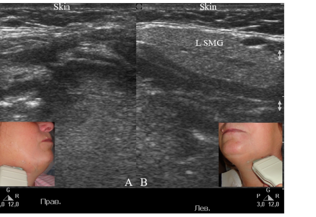
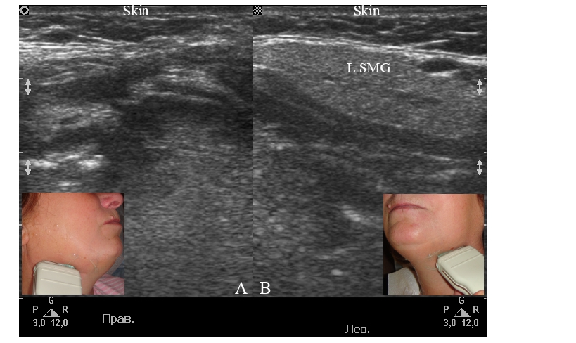
FIGURE 5. Comparison of gray scale sonograms of the right (A) and left (B) submandibular areas on the day 6 from the moment of the patient's hospitalization and on the day 3 from the moment of the sialolith removal. The absence of the right submandibular gland and the presence of an unchanged left submandibular gland (L SMG) are noted. The “depth” of ultrasonography is 4.0 cm.
DISCUSSION
A thorough review of recurrent sialoliths in a residual Wharton’s duct after sialoadenectomy in the relevant literature spanning the past 60 years was conducted by Su et al. (2025) [3].
According to Ellies et al. (1996), the shortest recorded time to symptoms of recurrent sialoliths in a residual duct is a month after surgery and a year after surgery [12]. The longest recorded period is thirteen years after the surgery (Patton, 1987) [5].
Interestingly, Koo et al. (2009) even report the possibility of sialolithiasis within the ipsilateral remaining Wharton’s duct in patients with isolated aplasia of a unilateral submandibular gland [13].
Su et al. (2025) emphasize new calculi may form again in the residual duct even if the submandibular glands were removed [3]. Kim et al. (2004) believe that such cases with recurrent sialolithiasis following submandibular gland excision should be treated by excising the sublingual glandular complex along with the remaining submandibular duct [7].
Douglas et al. (2021) emphasize that sialodocholith removal with duct remnant preservation may be possible, but with chronically inflamed deep lobes or the rare case of ductal premalignancy, excision of the residual submandibular gland and duct remnant may be needed [11].
Markiewicz et al. (2007) argue that treatment of sialolithiasis should be as conservative as possible [6]. Therefore, in the case of our patient, we followed exactly this tactic. And the incision was performed considering the anatomy of the sublingual cave [14, 15].
The shape of the removed sialolith in our case corresponds to the shape of calculi localized in the Wharton’s duct [2], and not to the calculi characteristic of the body of sublingual gland, in which they are predominantly round [16].
The fact that sialolith was not visualized on ultrasound may be explained by its location in the anterior part of the Wharton’s duct near its orifice. According to the literature, sialoliths in this area are the worst visualized on ultrasound. Thomas et al. (2017) prove that the majority of missed salivary stones were anterior Wharton's duct stones [17]. These sialoliths were likely missed due to an incomplete examination [17].
In our patient, it would theoretically be possible to consider the presence of a sialocele due to incomplete submandibular gland removal if the complication occurred in the immediate postoperative period and if there was no calculus. Benslimane et al. (2020), based on a systematic review and critical analysis of the evidence, stated that sialocele formation after the partial removal of the submaxillary gland for aesthetic indications was noted in 1.3% of cases [18]. Discussion of removal of the superficial aspect of the submandibular gland to improve the aesthetic appearance of the face and neck has been published in a numerous source [19, 20]. Therefore, although some surgeons may make a mistake in sialolithiasis and incompletely remove the deep lobe of the gland, in our case it was not possible to sufficiently prove from ultrasound data that a portion of the gland remained unremoved.
We consider it appropriate to quote the hypothesis of Ying et al. (2016) [8], namely: “Anatomically, a communication exists between the sublingual glandular complex and the submandibular gland duct. The sublingual gland communicates with the Wharton’s duct via the Bartholin’s duct or directly into the floor of the mouth. Therefore, the communication between the sublingual gland and the Wharton’s duct is hypothesized to provide an anatomical basis for sialolith formation. In addition, the sublingual gland is predominantly a mucus secreting gland, and the viscous saliva in the relatively stagnant environment within the residual part of the Wharton’s duct further facilitates calculus formation” [6, 21-23]. We could be more inclined to this theory if we had the patient’s examination data before the surgery 6 years ago and no radiological signs at that moment of sialolith in the right Wharton’s duct. We can only assume that in this case it is the type 2 of duct system pattern according to the classification of Zhang et al. (2010) [21, 22]. To understand sialodochitis of the Wharton’s duct and floor of mouth swelling in our patient, it is important to understand the histological structure of the duct, which consists of inner epithelium, basement membrane, and outer connective tissue carrying small blood vessels, capillaries, and autonomic nerve fibers [24]. Main duct features a two-layer columnar epithelium that eventually transitions into stratified squamous epithelium where it opens into the sublingual caruncle at the floor of the mouth.
Summing up this case, it is worth noting that ultrasound allowed us to reliably diagnose sialodochitis of the residual Wharton’s duct, analyze the condition of the tissues of the floor of mouth in the areas of both the removed gland and the left submandibular gland, and verify reactive lymphadenitis. Even though ultrasound the team failed to verify the calculus, assessment of the surrounding tissues is an essential diagnostic test that allowed us to exclude an odontogenic abscess.
CONCLUSION
The case presented in this article complements the diagnostic picture of the small number of reports of recurrent sialolithiasis following submandibular gland excision. And ultrasound images are the first in the English literature to demonstrate the state of floor of mouth tissues with this type of pathology.
DISCLOSURES
Author Contributions: Concept and Design – I.I.F.; Literature Search – O.S.C.; Writing – I.I.F., O.S.C.; Critical Review – O.Y.M.
Declaration of Interests: The authors have no conflicts of interest to declare.
Informed Consent: Written consent was obtained from patient for publication of photographs.
Fundings: No funding was received for this study.
Peer-Review: Externally peer-reviewed.
AI Statement: The authors declared that artificial intelligence was not used in this article.
REFERENCES (24)
-
Tymofieiev OO. Diseases of the salivary glands. VNTL-Klasyka; 2007. Ukrainian.
-
Tymofieiev OO. Non-neoplastic diseases of the salivary glands. In: Tymofieiev OO, ed. Surgical Dentistry and Maxillofacial Surgery. Vol 2. Marchenko TV; 2025:chap 2. Ukrainian.
-
Su LW, Sun H, Zhang HY, Wu Y. Large recurrent sialoliths in a residual Wharton duct after sialoadenectomy: Two case reports and literature review. Medicine (Baltimore). 2025;104(2):e41231. https://doi.org/10.1097/md.0000000000041231
-
Berini-Aytes L, Gay-Escoda C. Morbidity associated with removal of the submandibular gland. J Craniomaxillofac Surg. 1992;20(5):216-219. https://doi.org/10.1016/s1010-5182(05)80318-x
-
Patton DW. Recurrent calculus formation following removal of the submandibular salivary gland. Br J Oral Maxillofac Surg. 1987;25(1):15-20. https://doi.org/10.1016/0266-4356(87)90152-5
-
Markiewicz MR, Margarone JE 3rd, Tapia JL, Aguirre A. Sialolithiasis in a residual Wharton’s duct after excision of a submandibular salivary gland. J Laryngol Otol. 2007;121(2):182-185. https://doi.org/10.1017/s0022215106003525
-
Kim EA, Kim HS, Kim HK, Jung SM. Recurrent sialolithiasis on remnant Wharton’s duct following submandibular gland resection. Korean J Otolaryngol-Head Neck Surg. 2004;47(11):1185-1187.
-
Ying X, Kang J, Zhang F, Dong H. Recurrent sialoliths after excision of the bilateral submandibular glands for sialolithiasis treatment: A case report. Exp Ther Med. 2016;11(1):335-337. https://doi.org/10.3892/etm.2015.2849
-
Sivasubramaniam R, Choroomi S. Sialolithiasis in a remnant Wharton’s duct: a case study and discussion. J Otolaryngol ENT Res. 2015;2(1):51-53. https://doi.org/10.15406/joentr.2015.02.00012
-
Potash A, Hoffman HT. Retrograde sialendoscopy: a new technique for avoiding retained ductal stones. Ann Otol Rhinol Laryngol. 2012;121(1):38-43. https://doi.org/10.1177/000348941212100107
-
Douglas JE, Thomas WW, Wen CZ, et al. Sialendoscopy for sialodocholithiasis following submandibular gland excision: six variations on a theme. World J Otorhinolaryngol Head Neck Surg. 2021;7(4):291-295. https://doi.org/10.1016/j.wjorl.2020.10.003
-
Ellies M, Laskawi R, Arglebe C, Schott A. Surgical management of nonneoplastic diseases of the submandibular gland. A follow-up study. Int J Oral Maxillofac Surg. 1996;25(4):285-289. https://doi.org/10.1016/s0901-5027(06)80058-5
-
Koo BS, Lee SW, Lee YM, et al. Sialolithiasis in a stump of Wharton's duct of an aplastic unilateral submandibular gland. Int J Oral Maxillofac Surg. 2009;38(1):93-95. https://doi.org/10.1016/j.ijom.2008.11.008
-
Uz Z, Dilken O, Milstein DMJ, et al. Identifying a sublingual triangle as the ideal site for assessment of sublingual microcirculation. J Clin Monit Comput. 2023;37(2):639-649. https://doi.org/10.1007/s10877-022-00936-9
-
Bradley PJ. Sublingual gland. Operat Tech Otolaryngol Head Neck Surg. 2018;29(3):168-176. https://doi.org/10.1016/j.otot.2018.06.006
-
Hong KH, Yang YS. Sialolithiasis in the sublingual gland. J Laryngol Otol. 2003;117(11):900-902. https://doi.org/10.1258/002221503322542971
-
Thomas WW, Douglas JE, Rassekh CH. Accuracy of ultrasonography and computed tomography in the evaluation of patients undergoing sialendoscopy for sialolithiasis. Otolaryngol Head Neck Surg. 2017;156(5):834-839. https://doi.org/10.1177/0194599817696308
-
Benslimane F, Kleidona IA, Cintra HPL, Ghanem AM. Partial removal of the submaxillary gland for aesthetic indications: A systematic review and critical analysis of the evidence. Aesthetic Plast Surg. 2020;44(2):339-348. https://doi.org/10.1007/s00266-019-01546-8
-
Buckner H, Shah AR. Partial submandibular gland resection with ultrasonic dissector vs electrocautery. Aesthet Surg J Open Forum. 2025;7:ojaf011. https://doi.org/10.1093/asjof/ojaf011
-
Morel SBA, Macias LH, Chopra R, et al. Aesthetic submandibular gland resection: A review of complication incidence and prevention. Aesthet Surg J. 2025;45(9):869-874. https://doi.org/10.1093/asj/sjaf096
-
Zhang L, Xu H, Cai ZG, et al. Clinical and anatomic study on the ducts of the submandibular and sublingual glands. J Oral Maxillofac Surg. 2010;68(3):606-610. https://doi.org/10.1016/j.joms.2009.03.068
-
Nikulin DS, Fesenko II, Cherniak OS. Sialoliths in ipsilateral Bartholin’s and Wharton’s duct: the first clinical and ultrasound report. J Diagn Treat Oral Maxillofac Pathol. 2020;4(7):112-118. https://doi.org/10.23999/j.dtomp.2020.7.2
-
Capaccio P, Torretta S, Ottavian F, Sambataro G, Pignataro L. Modern management of obstructive salivary diseases. Acta Otorhinolaryngol Ital. 2007;27(4):161-172.
-
Chylinska W. Submandibular Gland (H&E Staining) - PUMS Histology Slides Review Series. Department of Histology and Embryology of Poznan University of Medical Sciences; 2026. Accessed January 11, 2026. https://www.youtube.com/watch?v=VtlV4Wm0q28&t=13s