July 31, 2019
https://doi.org/10.23999/j.dtomp.2019.7.2
J Diagn Treat Oral Maxillofac Pathol 2019;3:174–5.
Under a Creative Commons license
HOW TO CITE THIS ARTICLE
Demidov VH, Ripolovska OV. How multiple the submandubular gland sialoliths can be? J Diagn Treat Oral Maxillofac Pathol 2019;3(7):174–5.
A 52-year-old man presented to the Maxillofacial Surgery Department of Kyiv Regional Clinical Hospital with a several-year history of swelling in the right submandibular area and salivary colic during exacerbation. A physical examination showed significantly enlarged and firm right submandibular gland (Panel A, arrow).
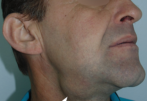
PANEL A. A physical examination showed significantly enlarged and firm right submandibular gland (arrow).
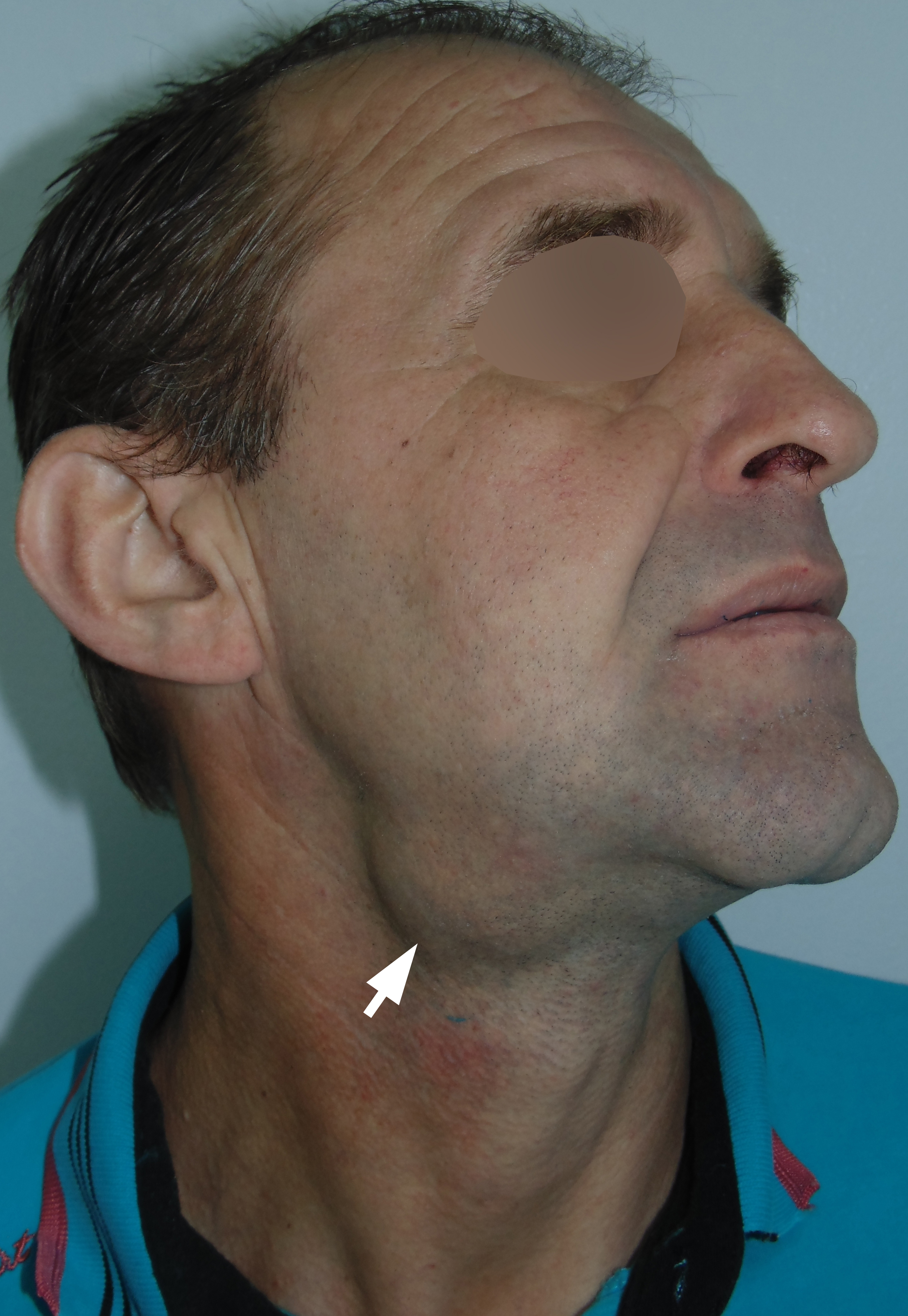
The gland was permanently increased in size during last months. Ultrasound (Panel B – Video) shows dilated intragandular ducts and multiple sialoliths (arrowheads), which visualized as hyperechoic bodies with artifact of acoustic shadowing). Replacement of glandular tissue with fibrous one was also noted.
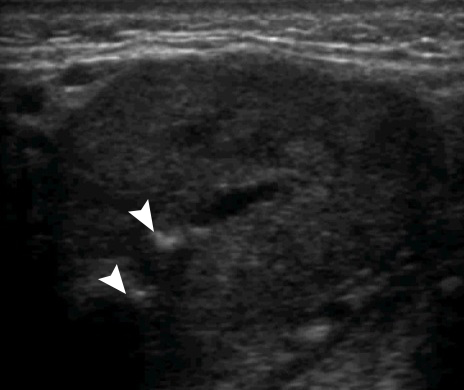
PANEL B – VIDEO. Ultrasound shows dilated intragandular ducts and multiple sialoliths (arrowheads), which visualized as hyperechoic bodies with artifact of acoustic shadowing.
VIDEO. Ultrasound shows dilated intragandular ducts and multiple sialoliths, which visualized as hyperechoic bodies with artifact of acoustic shadowing.
The patient underwent a complete gland removal and 8 different sized salivary stones have been found in the intraglandular duct system of the specimen (Panel C, arrows indicate sialoliths, and a fibrous tissue is indicated by asterisks).
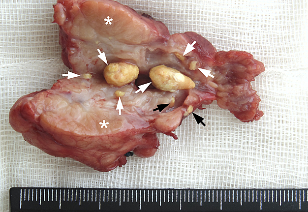
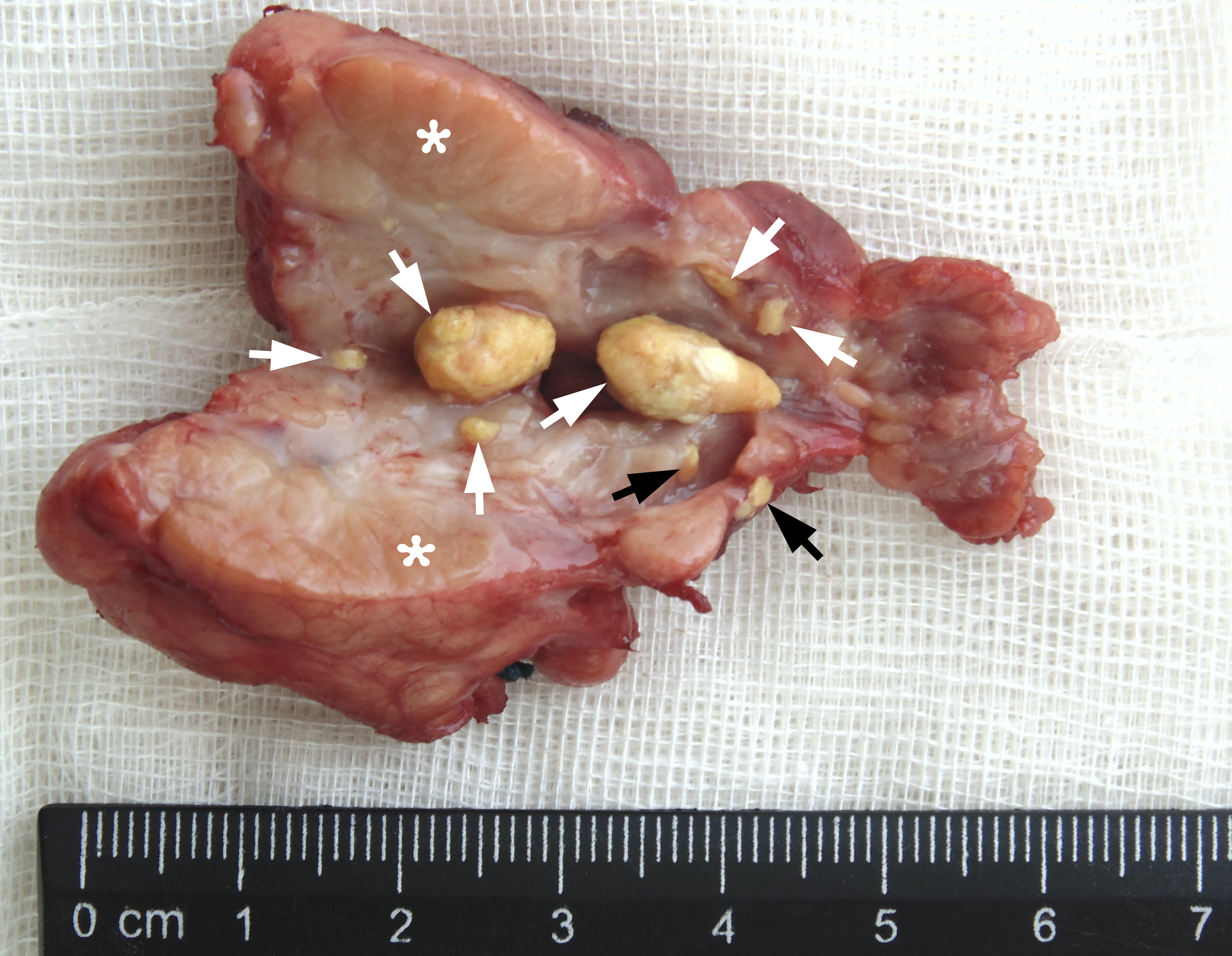
PANEL C. 8 different sized salivary stones have been found in the intraglandular duct system of the specimen (arrows indicate sialoliths, and a fibrous tissue is indicated by asterisks).
Two stones reached 8 and 10 mm in longitudinal size, and six another sialoliths measured no more than 3 mm. The two microsialoliths, with less than 1 mm in size, were also found. At follow-up 6 months after the surgery no complaints were noted. ■ DTJournal.org